 Neuromelanin (NM) is a dark pigment found in the brain which is structurally related to melanin.
It is a polymer of 5,6-dihydroxyindole monomers.
Neuromelanin is found in large quantities in catecholaminergic cells of the substantia nigra pars compacta and locus coeruleus, giving a dark color to the structures.
It gives specific brain sections, such as the substantia nigra or the locus coeruleus a distinct color.
Neuromelanin accumulates with aging, noticeably after the first 2–3 years of life.
Neuromelanin protects neurons in the substantia nigra from iron-induced oxidative stress.
It is a true melanin.
Neuromelanin is biosynthesized from L-DOPA, precursor to dopamine, by tyrosine hydroxylase and aromatic acid decarboxylase.
Synaptic vesicles and endosomes accumulate cytosolic dopamine and transport it to mitochondria where it is metabolized by monoamine oxidase.
Excess dopamine and DOPA molecules are oxidized by iron into dopaquinones and semiquinones which are then phagocytosed and are stored as neuromelanin.
Neuromelanin concentration increases with age.
Neuromelanin may have a role in neuroprotection
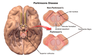
Neuromelanin-containing neurons in the substantia nigra undergo neurodegeneration during Parkinson’s disease.
Motor symptoms of Parkinson’s disease are caused by cell death in the substantia nigra, which may be partly due to oxidative stress.
This oxidation may be relieved by neuromelanin.
Patients with Parkinson’s disease have 50% the amount of neuromelanin in the substantia nigra as compared to similar patients of their same age, but without Parkinson’s.
The death of neuromelanin-containing neurons in the substantia nigra, pars compacta, and locus coeruleus have been linked to Parkinson’s disease and also have been visualized with neuromelanin imaging.
Neuromelanin has been shown to bind neurotoxic and toxic metals that could promote neurodegeneration.