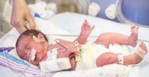
 Neonatal seizures are defined as seizures occurring within 4 weeks after birth in full-term infants or within 44 weeks of postmenstrual age in preterm infants.
The estimated incidence of neonatal seizures is 2.29 cases per 1000 live births.
Higher rates have been reported among preterm neonates than among full-term neonates (14.28 cases per 1000 vs. 1.10 per 1000).
Most neonatal seizures are transient in nature.
Most neonatal seizures are a result of acute metabolic disturbances, infectious processes, or acute focal cerebral lesions, and are considered to be provoked.
In full-term neonates, the most common cause of provoked seizures is hypoxic ischemic encephalopathy, followed in frequency by stroke and infection.
The most common cause is intraventricular hemorrhage in preterm neonates.
Identifying the provoking event in neonatal seizures is essential for determining management.
Provoked seizures are not considered to be epilepsy.
Provoked seizures typically do not require long-term treatment with antiseizure medication.
Neonatal epilepsy syndromes, which are uncommon, frequently have genetic causes, some of these syndromes require long-term treatment.
Neonatal seizures start focally but can spread to involve the entire brain.
Seizures that begin in a generalized fashion are rare.
Convulsive movements in babies are often complex, irregular, or subtle, and some seizures have only an electroencephalographic (EEG) component.
EEG is essential for the identification of neonatal seizures.
Clinical seizure types are usually associated with specific underlying causes:
Focal clonic seizures usually signify a cerebral infarction and the need for cranial imaging to confirm the diagnosis.
Tonic seizures are seen mainly with hypoxic ischemic encephalopathy but also with metabolic disorders, channelopathies, vascular disorders, or cortical malformations.
Myoclonic seizures, being sudden, brief, irregular limb contractions, metabolic disorders such as hypoglycemia or hyponatremia, amino acid disorders, or other inborn errors of metabolism are usually responsible.
Sequential seizures refers to the occurrence of more than one seizure type during an episode and are often genetically determined.
Epileptic spasms with sudden flexion, extension, or mixed flexion with extension of the proximal and truncal muscles also suggests genetic causes.
Autonomic seizures are characterized by alterations in cardiovascular, vasomotor, or respiratory patterns and are typically observed in neonates with hypoxic ischemic encephalopathy or intracranial hemorrhage.
Tremor, jitteriness, and some myoclonic movements can be mistaken for seizures.
Benign neonatal sleep myoclonus, neonatal hyperekplexia is a rare disorder of muscle rigidity, exaggerated startle reaction, and nocturnal myoclonus, with a normal EEG, and is also not an epilepsy syndrome.
The initial steps in managing neonatal seizures are to stabilize cardiovascular and respiratory function and then to identify the cause of the seizures.
Treatable abnormalities, such as hypoglycemia and electrolyte disorders can be rapidly detected and corrected, usually leading to cessation of seizures without the need for antiseizure medication.
EEG monitoring should be initiated as early as possible to establish the presence of seizures because some types of seizures tend to peak in incidence and severity within the first 24 hours, particularly those due to hypoxic ischemic encephalopathy.
Some seizures, such as those due to nonketotic hyperglycinemia, begin prenatally, and women may describe episodes of frequent, continuous, rhythmic jerking of the fetus.
Microcephaly may indicate cerebral dysgenesis, genetic abnormalities, or congenital infection.
Macrocephaly may be due to structural or genetic abnormalities;
Dysmorphic features suggest cerebral dysgenesis, often due to a genetic abnormality;
Neurocutaneous stigmata are indicative of specific disorders such as tuberous sclerosis or neurofibromatosis;
rash suggests infection or incontinentia pigmenti;
congenital heart disease is associated with perinatal stroke.
Workup for Acute Provoked Neonatal Seizures:
Screening for neonatal infection; toxicologic testing; metabolic testing for organic acidemias, urea cycle defects, and fatty acid oxidation defects, which may include amino acid levels, ammonia, lactate, pyruvate, very-long-chain fatty acids, urine, organic acids, biotinidase, pipecolic acid, and pyridoxal-5-phosphate, and examination of the placenta for pathological changes.
If an infectious cause is suspected, serum and cerebral spinal fluid cultures are generally obtained quickly and antimicrobial treatment is promptly initiated.
Continued seizures despite the administration of conventional antiseizure medication, a pyridoxine challenge may be attempted, for the rare condition known as pyridoxine-dependent developmental and epileptic encephalopathy.
Conventional 20-channel EEG or, amplitude-integrated EEG may be used to diagnose neonatal seizures.
Neuroimaging is essential in the detection of possible structural abnormalities in neonates with seizure.
Ultrasonography of the head is a first-line test because of its ease of use and accessibility.
Ultrasonographic assessment has high sensitivity (100%) and specificity (93.3%) for detecting intraventricular hemorrhages with ventricular enlargement, but the sensitivity is lower in the case of normal-size ventricles or small cerebellar or extraaxial hemorrhages.
Additional imaging with axial computed tomography or, preferably, magnetic resonance imaging (MRI) of the head can be performed.
Genetic testing is considered if there is no clear structural explanation for seizures, or if they are sequential seizures, epileptic spasms, or tonic seizures: epilepsy gene panels, chromosomal microarray, targeted gene testing, and whole-exome sequencing.
An increasing numbers of pathogenic variants have Ben identified as contributing to phenotypic epilepsy syndromes.
All neonatal seizures with both clinical and EEG correlates and those with only EEG evidence should be treated.
Medications used in the acute care setting are typically limited to intravenous formulations.
The International League Against Epilepsy (ILAE) recommends phenobarbital as the first-line antiseizure medication, regardless of the cause: hypoxic ischemic encephalopathy, stroke, hemorrhage, or genetic causes.
A randomized, controlled trial, showed that phenobarbital was more effective than levetiracetam at 24 hours for the treatment of neonatal seizures.
If the neonate does not respond to the first antiseizure medication, phenytoin, levetiracetam, midazolam, or lidocaine may be used as second-line intervention.
It is not clear regarding the best medication to be used after phenobarbital has failed to control the disorder: levetiracetam, and concise pyridoxine, pyridoxal phosphate, and folinic acid to correct uncommon vitamin-responsive epilepsies.
For term and near-term infants with moderate-to-severe hypoxic ischemic encephalopathy therapeutic hypothermia for 72 hours is now used routinely, to ameliorate the brain injury and improve later developmental outcomes.
Developmental and epileptic encephalopathies are defined by intractable seizures associated with developmental impairment or regression often due to an underlying cause: genetic, structural, or metabolic.
The self-limited neonatal epilepsy syndromes are due to pathogenic variants, begin between 2 and 7 days after birth and remits after 6 months.
Clues to this diagnosis are a family history of seizures and focal and tonic seizures at the onset of an episode.
Sodium-channel blockers are used when seizures are due to loss-of-function KCNQ2 and KCNQ3 variants.
Neuroimaging, genetic testing, and metabolic studies reveal the underlying cause in up to 80% of infants.
Treatment can be targeted at an underlying metabolic disorder, if present.
Surgical removal of a focal lesion is considered after the failure of two or more drug trials.
Some neonates may have coexisting movement disorders, cortical visual impairments, feeding difficulties, or orthopedic problems due to abnormal muscle tone and contractures.
KCNQ2-associated developmental and epileptic encephalopathy begins in the first few days of life, and is characterized mainly by focal tonic seizures.
Encephalopathy is present when the seizures begin.
Pyridoxine-dependent developmental and epileptic encephalopathy and pyridoxal phosphate deficiency–associated developmental and epileptic encephalopathy, are genetic syndromes because their treatment differs from that of other neonatal epilepsies.
With these disorders infants present within the first hours to days of life with encephalopathy and intractable seizures.
Seizures are frequent, often evolving into status epilepticus with focal or multifocal myoclonic movements of the face, arms, legs, and trunk; epileptic spasms may also occur.
CDKL5-associated developmental and epileptic encephalopathy, typically develops within the first few weeks of life, and the seizures are drug-resistant.
Neonates with this disorder typically present with hypotonia and sequential seizures with hyperkinetic, tonic epileptic spasms.
The prognosis for neonatal seizures varies with its cause, age at onset, seizure duration, and responsiveness to medication.
Self-limited epilepsy syndromes are characterized by frequent seizures, they remit spontaneously, with a typically good prognosis.
The developmental and epileptic encephalopathies, which are due to severe, diffuse brain injury, often with a genetic cause, and have a poor overall prognosis.
Untreated seizures can cause hippocampal sclerosis and worsen clinical outcomes.
Status epilepticus or seizures lasting longer than 12 to 13 minutes per hour are associated with a poor outcome, which is independent of the cause.
There is no definitive evidence that isolated seizures of brief duration have a negative outcome.
Animal suggest that the immature brain may be less susceptible to injury from seizures than the more mature brain.
Patterns on EEG can assist in determining the prognosis for neonates with clinically confirmed EEG seizures and for those with only EEG evidence of seizure activity.
Background burst suppression and ictal activity are associated with a poor prognosis.
