 Nemaline myopathy is also called rod myopathy or nemaline rod myopathy.
Nemaline myopathy affects less than 1 out of 50,000 on average.
At the electron microscopic level, rod-shaped components can often be seen in some of the muscle cells, and are diagnostic for the condition called nemaline rod myopathy.
The rods appear as a result of a dysfunctional muscle fiber.
There is no correlation between the number of rods found in the muscle cells and the amount of weakness a person has.
Gene mutations that lead to the nemaline myopathy are in genes that encode different components of the sarcomere.
It is a congenital, often hereditary neuromuscular disorder.
Characterized by muscle weakness, hypoventilation, swallowing dysfunction, and impaired speech ability.
Symptoms and severity change throughout one’s life to some extent.
The prevalence is estimated at 1 in 50,000 live births.
The risk of nemaline myopathy is the same in males and females.
Nemaline myopathy is the most common non-dystrophic myopathy.
Nemaline myopathy contains thread-like rods in muscle fibers, which are referred to as nemaline bodies.
The rods are diagnostic of the disorder.
Nemaline bodies are likely a byproduct of the disease process rather than a cause.
The disease is associated with delayed motor development or lack of motor development in severe cases.
There may be weakness in all skeletal muscles.
Weaknesses more profound in the proximal muscle groups than distal muscles.
The ocular muscles are usually uninvolved.
The disorder has a wide range of overlapping severity, from the most severe neonatal form which is incompatible with life, to a form so mild that it may not be diagnosed since the person appears to function at the lowest end of normal strength and breathing adequacy.
Respiratory problems are usually a major concern for people with all forms of NM, and respiratory infections are quite common.
NM shortens life expectancy in the more severe forms.
Proactive care allows most individuals to survive and even lead active lives.
Young children and babies lack movement and have a difficult time eating and breathing.
A sign of NM is a swollen face in disproportional areas.
Findings in newborns include swaying and a difficulty in moving, feeble muscles in the neck and upper rib cage area.
In adults, the most common symptom is respiratory problems, and mild to severe speech impediments.
Scoliosis is common.
Babies that have NM may take longer to walk than average due to the lack of muscle, or just muscle fatigue.
Elongated faces and a lower mandible are often observed in people with NM.
Muscle exhaustion generally appears between ages 20–50, however it
usually not progressive.
Gastroesophageal reflux, can be associated with NM.
Heart abnormalities rarely occur as a result of NM.
Most children with mild NM can eventually walk independently.
Some children use wheelchairs, walkers or braces, to enhance mobility.
With severe NM there is generally limited limb movement and use wheelchairs full-time.
Weakness in the trunk muscles, makes people with NM prone to scoliosis.
Scoliosis usually develops in childhood and worsens during puberty.
Patients may undergo spinal fusion surgery to straighten and stabilize their backs.
Early on patients often have hyper mobility in their joints.
Joint deformities and scoliosis subsequently occur.
Management of joint problems includes stretching exercises, physical therapy, braces, and surgery.
Vigorous exercise and the use of heavy weights are avoided.
Infants with severe NM frequently experience respiratory distress at or soon after birth.
Hypoventilation can begin insidiously, and it may cause serious health problems.
Throat muscle weakness is a main feature of nemaline myopathy.
Patients with the most severe forms of NM are unable to swallow and must receive their nutrition through feeding tubes.
Most people with intermediate and mild NM can take some or all of their nutrition orally.
Bulbar muscle impairment may impair communication.
Communicative skills may be improved with speech therapy, oral prosthetic devices, surgery, and augmentative communication devices.
Patients often have hypernasal speech as a result of poor closure of the area between the soft palate and the back of the throat.
NM is not associated with decreased cognition or intelligence.
Physical characteristics: weakness is usually in the proximal muscles, particularly respiratory, bulbar and trunk muscles.
Severe NM is clinically obvious at birth.
Patients with intermediate or mild NM may initially appear unaffected.
The physical abilities of a patient with NM do not correlate well either with genotype or with muscle pathology as observed in the biopsy.
Babies with NM are frequently observed to be floppy and hypotonic, but often gain strength as they grow.
The effect of muscle weakness on body features may become more evident with time.
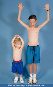
Adults with NM typically have a very slender physique.
Nemaline myopathy is caused by mutations in one of at least 11 different genes, and is a clinically and genetically heterogeneous disorder.
It has both autosomal dominant and autosomal recessive forms.
Diagnostic findings: muscle weakness, absent or low deep tendon reflexes, and a high-arched palate, along with electron-dense aggregates, called nemaline rods, being observed at the microscopic level within muscle fibers.
Genetic mutation provides confirmation:
The two most common gene mutations causing nemaline myopathy are found on NEB or ACTA1 genes.
NEB mutations usually result in symptoms present at birth or beginning in early childhood.
NEB mutations are autosomal recessive and account for 50% of affected nemaline myopathy patients.
Patients with NB mutations of NM are more affected in the muscles in their head, rather than their proximal muscles at the core of their body.
With this genetic mutation patients often cannot lift their heads and speak with a nasal voice, suggesting this kind of NM may lead to patients having higher intellect.
Occasionally with ACTA1 mutations with NM can be caused by an inheritance pattern of autosomal dominance, and results in about 15 to 25 percent of NM cases.
NM is also associated with de novo mutations in ACTA1, occurring spontaneously in the egg or sperm.
MYPN is the last found gene related to NM.
The Slow α-Tropomyosin Gene TPM3 and varies from case to case with its severity: affected people are weaker and more affected in their lower limbs than their upper limbs.
With nemaline myopathy, muscle contraction is adversely affected by altered pattern of the muscle fibers that make up the sarcomere so muscles are unable to contract efficiently or effectively.
Respiratory muscles are often more affected than other skeletal muscle groups.
Cardiac muscle is usually not affected, but patients may present with dilated cardiomyopathy.
The ocular muscles are usually not involved.
Diagnosis:
Electromyography
MRI of the Musculoskeletal System to confirm that muscle cells contain rod like structures.
Treatment:
Nemaline myopathy is not curable.
Use of stretches, exercise, braces and require care with neurologists, physical therapists, speech therapists and psychologists.
Many people live healthy active lives even with moderate to severe disease.
Some people have seen mild improvements in secretion handling, energy level, and physical functioning with supplemental L-tyrosine.
