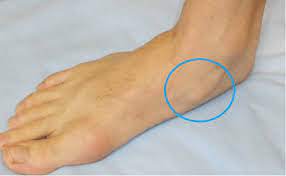
 Its location and impingement during foot strike predispose it to localized stress and remodeling.
The central area has relative avascularity.
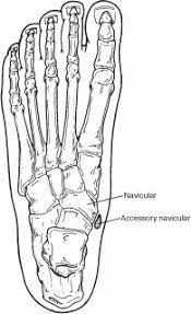
The navicular plays an important role in maintaining the medial longitudinally arch of the foot.
A disk-shaped bone that articulates distally with the 3 cuneiforms, proximally with the talar head, and, occasionally, laterally with the cuboid.
The distal articulation of the navicular with the 3 cuneiforms is by means of 3 facets that have a common synovial cavity. On the lateral side are the plantar, dorsal, and interosseous cuboideonavicular ligaments.
The blood supply is from small branches of the posterior tibial and dorsalis pedis arteries.
The medial and lateral areas of the navicular are relatively well vascularized compared with the central section of the navicular.
Essential for normal gait, from mid stance until push-off, as part of the medial longitudinal arch which is composed of the navicular, calcaneus, talus, 3 cuneiforms, and 3 medial metatarsals.
The transverse tarsal joint is essential for normal gait, as well, and is composed of the talonavicular joint and the calcaneocuboid joint.
This joint is flexible and plays an important role in absorbing ground impact and accommodating the foot to the ground.