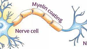
 Myelin is a specialized layer of membrane that surrounds and insulates the axons of neurons in the nervous system.
Myelin is a fatty substance that serves as an electrical insulator, enabling faster and more efficient transmission of nerve impulses along the axon.
Myelin is a lipid-rich material that surrounds nerve cell axons to insulate them and increase the rate at which electrical impulses (called action potentials) are passed along the axon.
Myelin sheaths the nerve in segments: in general, each axon is encased with multiple long myelinated sections with short gaps in between called nodes of Ranvier.
Myelin is formed in the central nervous system (CNS; brain, spinal cord and optic nerve) by glial cells called oligodendrocytes and in the peripheral nervous system (PNS) by glial cells called Schwann cells.
In the CNS, axons carry electrical signals from one nerve cell body to another.
In the PNS, axons carry signals to muscles and glands or from sensory organs such as the skin.
Each myelin sheath is formed by the concentric wrapping of an oligodendrocyte (CNS) or Schwann cell (PNS) process with a limb-like extension from the cell body, around the axon.
Myelin reduces the capacitance of the axonal membrane.
On a molecular level it increases the distance between extracellular and intracellular ions, reducing the accumulation of charges.
The structure of the myelin sheath is
discontinuous resulting in abrupt conduction, whereby the action potential jumps from one node of Ranvier, over a long myelinated stretch of the axon called the internode, before recharging at the next node of Ranvier, until it reaches the axon terminal.
Nodes of Ranvier are the short unmyelinated regions of the axon between adjacent long myelinated internodes.
At the axon terminal, this electrical signal provokes the release of a chemical message or neurotransmitter that binds to receptors on the adjacent post-synaptic cell, the nerve cell in the CNS or muscle cell in the PNS, at specialized regions called synapses.
Myelin’s insulin role is essential for normal motor functioning and sensory function and as demonstrated by the consequences of disorders that affect it, such as the genetically determined leukodystrophies;
Multiple sclerosis is an acquired inflammatory demyelinating disorder.
Inflammatory demyelinating peripheral neuropathies also exist.
Due to its high prevalence, multiple sclerosis, which specifically affects the central nervous system (brain, spinal cord and optic nerve), is the best known disorder of myelin.
In the CNS, oligodendrocyte progenitor cells (OPCs) differentiate into mature oligodendrocytes, which form myelin.
Myelination begins early in the 3rd trimester, with little myelin present in either the CNS or the PNS at the time of birth.
During infancy, myelination progresses rapidly, with increasing numbers of axons acquiring myelin sheaths, corresponding with the development of cognitive and motor skills, including language comprehension, speech acquisition, crawling and walking.
Myelination continues through adolescence and early adulthood and although largely complete at this time, myelin sheaths can be added in grey matter regions such as the cerebral cortex, throughout life.
Not all axons are myelinated.
In the PNS, a large proportion of axons are unmyelinated.
They are ensheathed by non-myelinating Schwann cells known as Remak Swann cells and arranged in Remak bundles.
In the CNS, non-myelinated axons intermingle with myelinated ones and are entwined, at least partially, by the processes of another type of glial cell the astrocyte.
CNS myelin differs slightly in composition and configuration from PNS myelin, but both perform an insulating function.
Being rich in lipid, myelin appears white, hence the name given to the “white matter” of the CNS.
Both CNS white matter tracts, such as the optic nerve, corticospinal tract and corpus callosum, and the PNS nerves, suv=ch as the sciatic nerve and the auditory nerve, which also appear white, each comprise thousands to millions of axons, largely aligned in parallel.
Blood vessels provide oxygen and energy substrates such as glucose to reach these fiber tracts.
These fiber tracts also contain other cell types including astrocytes and microglia in the CNS and macrophages in the PNS.
Myelin comprises approximately 40% water; the dry mass comprises between 60% and 75% lipid and between 15% and 25% protein.
The primary lipid of myelin is a glycolipid called galactocerebroside.
The intertwining hydrocarbon chains of sphingomyelin strengthen the myelin sheath.
Cholesterol is an essential lipid component of myelin, without which myelin fails to form.
Action potential propagation in myelinated neurons is faster than in unmyelinated neurons because of saltatory conduction.
Myelin’s main purpose is to increase the speed at which electrical action potentials propagate along the myelinated fiber.
In unmyelinated fibers, action potentials travel as continuous waves, but, in myelinated fibers, they are propagate by saltatory conduction.
The latter is markedly faster than the former, at least for axons over a certain diameter.
Myelin decreases capacitance and increases electrical resistance across the axonal membrane, and that permits larger body size by maintaining agile communication between distant body parts.
Myelinated fibers lack voltage-gated sodium channels along the myelinated internodes, exposing them only at the nodes of Ranvier.
Positively charged sodium ions can enter the axon through these voltage-gated channels, leading to depolarization of the membrane potential at the node of Ranvier.
The resting membrane potential is then rapidly restored due to positively charged potassium ions leaving the axon through potassium channels.
The sodium ions inside the axon then diffuse rapidly through the axonal cytoplasm to the adjacent myelinated internode and ultimately to the next node of Ranvier, triggering the opening of the voltage gated sodium channels and entry of sodium ions at this site.
Nodes of Ranvier have to be relatively closely spaced, to secure action potential propagation.
The action potential rechargesat consecutive nodes of Ranvier.
Along the myelinated internode, energy-dependent sodium/potassium pumps pump the sodium ions back out of the axon and potassium ions back into the axon to restore the balance of ions between the intracellular and extracellular fluids.
The myelination modifies the underlying axon by promoting the phosphorylation of neurofilaments, thus increasing the diameter or thickness of the axon at the internodal regions.
It helps cluster molecules of voltage-gated sodium channels at the node of Ranvier.
Myelination modulates the transport of cytoskeletal structures and organelles such as mitochondria, along the axon.
The myelinating cell fuels the axon, which uses a great deal of energy to restore the normal balance of ions between it and its environment, following the generation of action potentials.
When a peripheral fiber is severed, the myelin sheath provides a track along which regrowth can occur.
However, the myelin track layer does not ensure a perfect regeneration of the nerve fiber, so nerve fibers may find the correct muscle fibers, and some damaged motor neurons of the peripheral nervous system die without regrowth, resulting functional impairment.
Unmyelinated fibers and myelinated axons of the mammalian central nervous system do not regenerate.
Myelin production to form is stimulated by electrical activity in neurons that causes them to release ATP.
This ATP causes astrocytes to secrete a cytokine regulatory protein that promotes the myelinating activity of oligodendrocytes,
suggesting that astrocytes have an executive-coordinating role in the brain.
Demyelination, the loss of the myelin sheath insulating the nerves is the hallmark of some neurodegenerative autoimmune disease:, multiple sclerosis, acute disseminated encephalomyelitis, neuromyelitis optica, transverse myelitis, chronic inflammatory demyelinating polyneuropathy, Guillain–Barré syndrome, central pontine myelinosis, inherited demyelinating diseases such as leukodystrophy, and Charcot–Marie–Tooth disease.
Sufferers of pernicious anaemia can also suffer nerve damage of this type: Subacute combined degeneration of spinal cord can lead to slight peripheral nerve damage to severe damage to the central nervous system, affecting speech, balance, and cognitive awareness.
When myelin degrades, conduction of signals along the nerve can be impaired or lost, and the nerve eventually withers.
The immune system may play a role in demyelination associated with such diseases, including inflammation causing demyelination by overproduction of cytokines via upregulation of tumor necrosis factor
or interferon.
MRI evidence that docosahexaenoic acid DHA ethyl ester improves myelination in generalized peroxisomal disorders.
Demyelination results in diverse symptoms determined by the functions of the affected neurons as it disrupts signals between the brain and other parts of the body.
Typical symptoms of demyelination include: blurriness in the central visual field that affects only one eye, may be accompanied by pain upon eye movement, double vision, loss of vision/hearing, odd sensation in legs, arms, chest, or face, such as tingling or numbness. weakness of arms or legs, cognitive disruption, including speech impairment and memory loss, heat sensitivity, loss of dexterity, difficulty coordinating movement or balance disorder, difficulty controlling bowel movements or urination, fatigue, and tinnitus.
Cholinergic treatments, such as acetylcholinesterase inhibitors may have beneficial effects on myelination, myelin repair, and myelin integrity.
Increasing oligodendrocyte cholinergic stimulation, AChEIs, and other cholinergic treatments, such as nicotine, possibly could promote myelination during development and myelin repair in older age.
Cholesterol is a necessary nutrient for the myelin sheath, along with vitamin B12.
Dysmyelination is characterized by a defective structure and function of myelin sheaths; unlike demyelination, it does not produce lesions.
Such defective sheaths often arise from genetic mutations affecting the biosynthesis and formation of myelin.
Diseases where dysmyelination has been implicated include leukodystrophies (Pelizaeus–Merzbacher disease, Canavan disease, phenylketonuria) and schizophrenia.
Myelination is a critical process during development, and it continues throughout life in some regions of the brain.
Myelination is only prevalent in a few brain regions at birth and continues into adulthood.
Myelination process is not complete until about 25–30 years of age.
Myelination is an important component of intelligence, and white matter quantity may be positively correlated with IQ test results in children.
The myelin sheath is formed by specialized cells called oligodendrocytes in the central nervous system and Schwann cells in the peripheral nervous system.
Damage to the myelin sheath can disrupt nerve conduction and is associated with many neurological disorders, including multiple sclerosis, leukodystrophies, and peripheral neuropathies.