
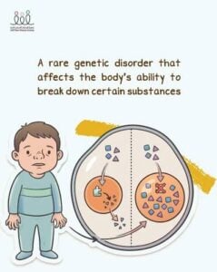
 Mucopolysaccharidoses (MPS) are a group of inherited lysosomal storage disorders caused by deficiency of one of the specific lysosomal enzymes required for the stepwise degradation of glycosaminoglycans (GAGs, formerly “mucopolysaccharides”).
The resulting intralysosomal accumulation of undegraded or partially degraded GAGs (heparan, dermatan, keratan, and chondroitin sulfate) causes progressive, multisystem cellular and organ damage, with excess GAG also excreted in the urine.
There are 11 known enzyme deficiencies giving rise to seven classically recognized MPS types (I, II, III, IV, VI, VII, IX), several with subtypes.
All are inherited autosomal recessively except MPS II (Hunter syndrome), which is X-linked recessive.
Overall incidence is approximately 1:25,000 births, with substantial regional variation (MPS I and III predominate in Europe.
MPS II predominates in East Asia.
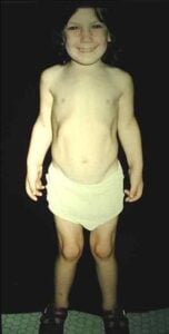
Shared clinical features commonly include coarse facial features, dysostosis multiplex, joint stiffness/contractures, short stature, hepatosplenomegaly, umbilical and inguinal hernias, hearing loss, corneal clouding, valvular heart disease and cardiomyopathy, obstructive and restrictive respiratory disease with sleep apnea, hydrocephalus, and cervical cord compression from odontoid dysplasia/dural thickening.
Neurocognitive involvement with developmental delay, regression, behavioral disturbance, is characteristic of the heparan-sulfate–accumulating types (I, II, III, VII), whereas MPS IV and VI generally spare cognition.
Manifestations are progressive, and initial signs are often subtle or nonspecific, contributing to diagnostic delay.
Clinical presentations overlap substantially among MPS I, II, and VI while MPS III shows a distinctly neurologic signature and MPS IV a distinctly skeletal one, molecular/enzymatic confirmation is essential.
The following clustering heatmap from a large re-diagnosis cohort illustrates these phenotypic signatures across subtypes.
Diagnosis proceeds on a stepwise basis:
clinical suspicion (particularly early non-inflammatory joint involvement, dysostosis multiplex, claw hand, or spinal deformity should prompt referral) for quantitative and qualitative urinary GAG analysis → identification of the predominant GAG to direct enzyme assay in leukocytes/fibroblasts → confirmatory molecular genetic testing.
Urinary GAG screening can yield false negatives, so a strong clinical suspicion warrants enzymatic/molecular confirmation regardless.
Molecular testing additionally aids prognosis, carrier screening, and prenatal/preimplantation diagnosis.
Treatment depends on the specific type and is best delivered by a multidisciplinary team with regular comprehensive surveillance.
Two disease-modifying modalities are established:
Enzyme replacement therapy (ERT): Approved options include laronidase (MPS I), idursulfase (MPS II), galsulfase (MPS VI, Naglazyme), and elosulfase alfa (MPS IVA); ERT for MPS VII is also available.
ERT improves somatic manifestations (hepatosplenomegaly, walking/respiratory capacity, joint mobility) but does not cross the blood-brain barrier and therefore does not control CNS disease; anti-drug antibodies and lifelong infusion burden are limitations.
Hematopoietic stem cell transplantation (HSCT): The standard of care for severe (neuronopathic) MPS I when performed early—ideally before 2.5 years of age—because donor-derived cells provide an endogenous, CNS-penetrating enzyme source that can preserve cognition and prolong survival.
HSCT has been ineffective in MPS II, largely attributed to late diagnosis, and skeletal and some cognitive abnormalities persist and progress even after successful engraftment.
Emerging therapies address the CNS and skeletal manifestations uncontrolled by current treatment.
Blood-brain-barrier–penetrant/fusion-protein ERT (e.g., pabinafusp alfa for MPS II CNS disease), intrathecal ERT, substrate reduction therapy, ex vivo hematopoietic stem-cell gene therapy (in Hurler syndrome), and in vivo gene therapy.
For types and phenotypes without disease-modifying options, supportive and palliative care—physical/occupational therapy, surgical intervention (e.g., cervical decompression, hernia repair, valve surgery), and symptom-directed medication—remains central.