 Specialized macrophages that protect the CNS from injury.
Act as the immune cells of the CNS.
Microglia are the most abundant immune cells in the brain and are an important defense against CNS injury.
Microglia have the ability to promote recovery mechanism with anti-inflammatory factors, and ability to inhibit large pro-inflammatory cytokines.
Undergo hyperplasia and hypertrophy during nociceptor release of neuropeptides and excitatory amino acids within the dorsal horn.
Over time, however, chronic inflammation causes the degradation of tissue and of the blood–brain barrier.
During this time, microglia generate reactive oxygen species and release signals to recruit peripheral immune cells for an inflammatory response.
Astrocytes are glial cells that are the most abundant cells in the brain.
They are involved in maintenance and support of neurons and compose a significant component of the blood–brain barrier.
After insult to the brain, such as traumatic brain injury, astrocytes may become activated in response to signals released by injured neurons or activated microglia.
Once activated, astrocytes may release various growth factors and undergo morphological changes.
Discharges prostanoids, cytokines and free radicals sensitizing the CNS to pain transmitting neurons.
Microglia actively survey their CNS environment and respond to neural injury.
Microglia actively survey their environment and change their cell morphology significantly in response to neural injury.
Acute inflammation in the brain is typically characterized by rapid activation of microglia.
During this period, there is no peripheral immune response.
Over time, however, chronic inflammation causes the degradation of tissue and of the blood–brain barrier.
During this time, microglia generate reactive oxygen species and release signals to recruit peripheral immune cells for an inflammatory response.
Astrocytes are glial cells that are the most abundant cells in the brain.
They are involved in maintenance and support of neurons and compose a significant component of the blood–brain barrier.
Acute inflammation in the brain is typically characterized by rapid activation of microglia.
Microglia are a type of neuroglia located throughout the brain and spinal cord.
Microglia account for about 10-15% of cells found within the brain.
Microglia are the resident macrophage cells, they act as the first and main form of active immune defense in the central nervous system (CNS).
Microglia and other neuroglia including astrocytes are distributed in large non-overlapping regions throughout the CNS.
Microglia are key cells in overall brain maintenance.
Microglia are constantly scavenging the CNS for plaques, damaged or unnecessary neurons and synapses, and infectious agents.
Microglia are extremely sensitive to even small pathological changes in the CNS, that are achieved in part by the presence of unique potassium channels that respond to even small changes in extracellular potassium.
Microglia are also key players in the sustainment of normal brain functions under healthy conditions, constantly monitor neuronal functions through direct somatic contacts and exert neuroprotective effects when needed, play important roles in maintaining brain function and responding to injury and inflammation.
They represent the resident immune system of the CNS.
Microglia have many functions, including:
1. Immune response: Like other immune cells, microglia actively participate in the immune response against pathogens, toxins, and other damaging agents in the CNS.
Microglia can engulf and eliminate foreign molecules, dead cells, and debris in the brain.
2. Inflammation: Microglia release cytokines and other inflammatory molecules that can exacerbate neuroinflammation, which can in turn contribute to the progression of neurodegenerative diseases.
3. Tissue remodeling: Microglia can change shape and position themselves to help shape the brain’s structure and connections.
4. Neuroprotection: Microglia release neurotrophic factors that promote neuron growth and survival.
Microglia may play important roles in brain development and various aspects of brain function from learning to complex behavior.
Microglial overactivation can contribute to damage and nerve cell death: Alzheimer’s disease, Parkinson’s disease, and stroke.
The brain and spinal cord are not usually accessed directly by pathogenic factors in the body’s circulation due to a series of endothelial cells known as the blood–brain barrier (BBB)>
The BBB prevents most infections from reaching the vulnerable nervous tissue.
If infectious agents are directly introduced to the brain or cross the blood–brain barrier, microglial cells must react quickly to decrease inflammation and destroy the infectious agents.
Because few antibodies are small enough to cross the blood–brain barrier,
Microglia must be able to recognize foreign bodies, swallow them, and act as antigen-presenting cells activating T-cells.
Microglial cells are extremely plastic, and undergo structural changes based on location and system needs so as to be able to fulfill the vast variety of functions that microglia perform.
They adopt a specific form, or phenotype, in response to the local conditions and chemical signals they have detected.
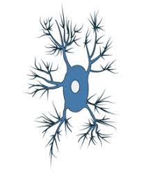
The resting form of microglia is composed of long branching processes and a small cellular body.
The cell body of the ramified form remains in place while its branches are constantly moving and surveying the surrounding area.
Unlike activated or ameboid microglia, ramified microglia do not phagocytose cells and secrete fewer immunomolecules.
Microglia in this state are able to search for and identify immune threats while maintaining homeostasis in the CNS.
In the resting state microglia are active in chemically surveying the environment.
Ramified microglia can be transformed into the activated form in response to injury or threat.
Microglia can be activated by a variety of factors including: pro-inflammatory cytokines, cell necrosis factors, lipopolysaccharide, and changes in extracellular potassium as indicative of ruptured cells.
Activated microglia cells undergo morphological changes including the thickening and retraction of branches, uptake of MHC proteins, expression of immunomolecules, secretion of cytotoxic factors, secretion of recruitment molecules, and secretion of pro-inflammatory signaling molecules.
Microglia also undergo rapid proliferation in order to increase their numbers.
Activated phagocytic microglia are the immune-responsive form of microglia.
Activated phagocytic microglia take on a large, ameboid shape.
Activated phagocytic microglia have the antigen presenting, cytotoxic and inflammation-mediating signaling of activated non-phagocytic microglia, they are also able to phagocytize foreign materials and display the resulting immunomolecules for T-cell activation.
Phagocytic microglia travel to the site of the injury, engulf the offending material, and secrete pro-inflammatory factors to promote more cells to proliferate and do the same.
Activated phagocytic microglia also interact with astrocytes and neural cells to fight off any infection or inflammation as quickly as possible with minimal damage to healthy brain cells.
Activated phagocytic microglia have free movement throughout the neural tissue, which allows it to fulfill its role as a scavenger cell.
Amoeboid microglia are able to phagocytose debris, but do not fulfill the same antigen-presenting and inflammatory roles as activated microglia.
Amoeboid microglia are especially prevalent during the development and rewiring of the brain, when there are large amounts of extracellular debris and apoptotic cells to remove.
This form of microglial cell is found mainly within the perinatal white matter areas in the corpus callosum.
Gitter cells are the eventual result of microglial cells’ phagocytosis of infectious material or cellular debris.
After engulfing a certain amount of material, the phagocytic microglial cell becomes unable to phagocytose any further materials, resulting in a cellular mass is known as a granular corpuscle.
By looking at tissue stained to reveal gitter cells, pathologists can visualize healed areas post-infection.
Perivascular microglia are mainly found encased within the walls of the basal lamina.
Perivascular microglia perform normal microglial functions, but are replaced by bone marrow-derived precursor cells on a regular basis.
They express MHC class II antigens, and react strongly to macrophage differentiation antigens.
Microglia are essential for the repair of vascular walls, as perivascular microglia promote endothelial cell proliferation, allowing new vessels to be formed and damaged vessels to be repaired.
Juxtavascular microglia have direct contact with the basal lamina wall of blood vessels but are not found within the walls.
Juxtavascular microglia express MHC class II proteins even at low levels of inflammatory cytokine activity.
Juxtavascular microglia do not exhibit rapid turnover or replacement with myeloid precursor cells on a regular basis.