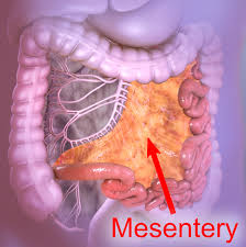
Refers to a continuous set of tissues that attaches the intestines to the abdominal wall in humans and is formed by the double fold of peritoneum.
It stores fat and allows blood vessels, lymphatics, and nerves to supply the intestines.
The mesocolon is a single structure, deriving from the duodenojejunal flexure and extending to the distal mesorectum layer.
The mesentery of the small intestine arises from the root of the mesentery.
The root of the mesentery extends from the duodenojejunal flexure to the ileocaecal junction, is about 15 cm long, 20 cm in width, and is directed obliquely from the duodenojejunal flexure at the left side of the second lumbar vertebra to the right sacroiliac joint.
The root of the mesentery lies behind the transverse colon and the greater omentum.
The mesentery is attached to the colon at the gastrointestinal margin and continues as the mesocolon.
The parts of the mesocolon are named from the part of the colon to which they attached: transverse mesocolon attaching to the transverse colon, the sigmoid mesocolon attaching to the sigmoid colon, the mesoappendix attaching to the appendix, and the mesorectum attaching to the upper third of the rectum.
The mesocolon is continuous from the ileocaecal to the rectosigmoid level.
A mesenteric confluence occurs at the ileocaecal and rectosigmoid junctions, and at the hepatic and splenic flexures.
Confluences involve peritoneal and omental attachments.
The proximal rectum originates at the confluence of the mesorectum and mesosigmoid.
The ileocaecal flexure arises at the point where the ileum is continuous with the caecum around the ileocaecal mesenteric flexure.
The hepatic flexure is formed between the right mesocolon and transverse mesocolon at the mesenteric confluence.
The colonic component of the hepatic flexure is draped around this mesenteric confluence.
The splenic flexure is formed by the mesenteric confluence between the transverse and left mesocolon.
A continuous peritoneal fold, at every flexure, lies outside the colonic/mesocolic complex tethering it to the posterior abdominal wall.
The transverse mesocolon is attached to the transverse colon that lies between the colic flexures.
The sigmoid mesocolon is that region of the mesentery to which the sigmoid colon is attached at the gastrointestinal mesenteric margin.
The mesoappendix is the portion of the mesocolon connecting the ileum to the appendix. It may extend to the tip of the appendix. It encloses the appendicular artery and vein, as well as lymphatic vessels, nerves, and often a lymph node.
The mesorectum is that part attached to the upper third of the rectum.
Adhesions frequently tether the greater omentum to the cephalad aspect of the transverse mesocolon.
The mesentery has a surface mesothelium and underlying connective tissue.
Adipocytes lobules within the body of the mesocolon are separated by fibrous septae arising from connective tissue.
Two mesothelial layers separate the mesocolon and underlying retroperitoneum.
The gastrointestinal tract and associated dorsal mesentery are subdivided into foregut, midgut, and hindgut regions based on the respective blood supply.
The portion of the dorsal mesentery that attaches to the greater curvature of the stomach, is known as the dorsal mesogastrium.
The part of the dorsal mesentery that suspends the colon is termed the mesocolon.
The dorsal mesogastrium develops into the greater omentum.
The part of the ventral mesentery that attaches to the stomach is known as the ventral mesogastrium.
Thinning of the mesoderm or ventral mesogastrium forms the lesser omentum, which attaches the stomach and duodenum to the anterior abdominal wall.
This leaf of mesoderm is divided into two parts – the lesser omentum between the stomach and liver, and the falciform and coronary ligaments between the liver and the abdominal wall and diaphragm.
In Crohn’s disease , the mesentery is frequently thickened.
Thrombosis of the superior mesenteric vein can cause mesenteric ischemia also known as ischemic bowel.
A volvulus, a twisted loop of the small intestine that also can enclose the mesentery too tightly can cause ischemia.
The total mesocolic excision (TME) operation has become the standard for the management of rectal cancer.
Recently, the surgical principles underpinning TME in rectal cancer have been extrapolated to colonic surgery, and it reduces local five-year recurrence rates in colon cancer from 6.5% to 3.6%, while cancer-related five-year survival rates in patients resected for cure increased from 82.1% to 89.1%.
There is a rich lymphatic network embedded within the mesenteric connective tissue.