 Median sternotomy is a type of surgical procedure in which a vertical inline incision is made along the sternum, after which the sternum itself is divided using a sternal saw.
The procedure provides access to the heart and lungs for surgical procedures such as heart transplant, lung transplant, corrective surgery for congenital heart defects, or coronary artery bypass surgery.
Indications: cardiac surgery, allows access to both pleural spaces, making it useful for some thoracic operations, retrosternal goiter access and esophagectomy.
Contraindications: previous surgery using the median sternotomy is associated with an increased risk of perioperative complications, obesity, COPD, radiation therapy to the chest, and cardiomegaly.
In cardiac surgery, a 5% risk exists of bleeding or tamponade following a first cardiac operation.
Emergency re-exploration is required for excessive mediastinal bleeding.
General anesthesia is dependent upon the type of operation to be performed: single lung ventilation is required, patients should be intubated with a double lumen endotracheal tube.
Thebrisk of hemorrhage is greater with redo sternotomy compared to the initial sternotomy.
For first-time operations, a standard pneumatic sternal saw is used.
In redo and pediatric surgery an oscillating saw is used.
The patient should be in the supine position
A roll os positioned in the interscapular region in order to improve access to the sternum by extending the neck and elevating the sternal notch.
A skin incision is made with a scalpel that extends from the midpoint between the angle of Louis and the sternal notch to below the xiphoid process.
An emergency sternotomy is generally performed for hemorrhage or tamponade following cardiac surgery.
Redo sternotomy is associated with a higher risk of complications then an initial sternotomy.
The standard procedure for closing the sternal incision is to place wire sutures to secure the sternum.
In neonatal cardiac surgery, the sternum is occasionally left open for 24-72 hours if there is a high risk of tamponade.
Recently the use of titanium plate fixation facilitates sternal stability in select patients.
The use of kryptonite bone cement which is a biocompatible polymer which can be used to adhere the bone edges.
Complications of sternotomy:
wound infection and dehiscence.
Deep sternal wound infection (DSWI)
occurs in 1-4% of cardiac surgery patients, although the incidence is increased with advanced patient age, diabetes, obesity, smoking, steroid therapy, and COPD.
Prolonged surgery or reopening of the sternotomy also puts the patient at higher risk of DSWI.
Chest hair should be shaved prior to surgery, and prophylactic antibiotics should be administered.
The incidence or DSWI is reduced by preoperative chlorhexidine gluconate showers and nasal mupirocin the night prior to and the morning of surgery.
Deep sternal wound infections are often associated with fever, leukocytosis, a sternal click with movement or respiration, purulent discharge from the sternal wound, and sternal dehiscence.
Treatment is with intravenous antibiotics and analgesia.
Superficial wound infection has an incidence of around 3-10% and is more common than deep sternal wound infections.
Massive hemorrhage is a major risk in a redo sternotomy; adhesions from the can result in major structures being attached to the chest wall.
Redo sternotomy carries a 1-3% risk of right ventricular trauma, which is associated with a 50% mortality rate.
Trauma to a patent coronary graft may occur.
Accidental trauma can also occur to the right atrium with redo sternotomy.
Preoperatively antiplatelet medications and antithrombotic agents should be discontinued several days prior to the sternotomy.
Sympathetic stimulation that occurs with sternotomy may result in tachyarrhythmias.
In redo surgery, damage to previous grafts is a risk, and cardiac manipulation may cause dysrhythmias including intractable ventricular fibrillation.
Sternal instability and pseudoarthrosis occurs 1-2% of patients following sternotomy.
Features of sternal instability include costochondral pain, clicking sensation on movement, and palpable instability on coughing.
Brachial plexus injury may result from sternal retraction.
Treatment is with analgesia and physiotherapy.
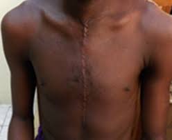
Hypertrophic keloid scarring is common with midline incisions.
Keloid scars are especially likely to develop in patients of African descent.