 ARMD age related macular degeneration.
A progressive retinal disorder affecting the macula, the central portion of the retina responsible for sharp vision.
Leading cause of irreversible visual impairment and blindness in the U.S.
AMD is the most common cause of incurable blindness IN older adults.
Geographic atrophy is an advanced stage of dry AMD in leads to progressive, irreversible death of a photoreceptors and retinalpigment, epithelial cells, causing profound vision loss.
Early and intermediate stages of AMD may not cause symptoms, late stage AMD can lead to severe vision impairment, interfering with visual acuity, such as reading and recognizing faces.
Early symptoms of AMD is associated with difficulty performing tasks on the low play conditions and low contrast surroundings.
About 40% of patients seek assistance due to visual distortion, approximately 38% reported decline in vision characterized by blurred vision, loss of visual acuity or difficulty focusing.
In industrialized countries age related macular degeneration (AMD) is a leading cause of blindness in adult older than 60 years of age.
Constitutes 6 to 9% of global legal blindness.
The global cost of visual impairment from AMD is more than $300 billion, including more than $250 billion in direct healthcare costs.
The estimated global prevalence of AMD is 8.7% and it is estimated that more than 196 million persons worldwide and more than 20 million people in the US have AMD.
In 2019 an estimated 20 million people in the US were living with AMD,and approximately 1.5 million of these had late stage AMD.
AMD is more common in individuals of European, and North American ancestry than in those of Asian, Hispanic, or African ancestry.
Vision loss from AMD is most prevalent in the white population.
It is suspected that this racial bias is related to the protection of ocular pigmentation.
GPR 143 (G protein receptor) a ligand for levodopa, which is an intermediate product of pigment synthesis and it is likely that retinal pigment epithelium is the primary tissue that initiates AMD pathobiology.
The incidence of AMD is similar among men and women.
With early or intermediate AMD patients may be asymptomatic.
Symptomatic patients present with blurred or decrease vision in one or both eyes, distortion, blind spots in or around a central vision, and difficulty with visual function and daily activities such as reading and driving, especially in poorly lit settings.
It primarily affects the macula, which is the cone photo receptor-rich central part of the retina.
It is characterized by a gradual loss of the retinal pigment epithelium and consequent photoreceptor death in the macula, resulting in patchy, wavy central vision and eventual blindness.
Neural, structural and vascular layers undergo degenerative changes, leading to cell death, including photoreceptors, which are specialized neurons that convert light into electrical signals that are processed in the visual cortex to form images: The retinal pigment epithelium, a cell layer that is adjacent to the photoreceptors, and the Bruch membrane, a collagenous layer that supports and separates the retinal pigment epithelium from the choroid, and the choroidcapillaris, which is a fine net mesh work of capillaries located in the innermost aspect of the choroid.
It is considered disease on a continuum of three clinical stage is encompassing early signs of retinal degeneration to more obvious features without overt visual symptoms and subsequently late stage manifestations, the latter accompanied by severe vision loss.
Most common cause of severe vision loss in elderly persons in developed countries and accounts for one-third of cases of untreatable vision loss.
Increasing age is its strongest risk factor, and with exponential growth of the aging population, it is suggested global prevalence will increase to 288 million persons in 2040.
Late stage AMD is associated with an incidence of 0.31 per thousand in those age 55 to 59 years and up to 36.7 per thousand in those age 90 years or older.
The estimated heritability of late stage AMD is approximately 71%, indicating that approximately 71% of the risk of AMD is genetic.
Genome wide association studies identified CFH and ARMS2 – HTRA1 that are cigstrongly linked to AMD.
A painless, irreversible, degenerative eye condition associated with the damage and ultimate death of photoreceptors.
AMD is multifactorial related to: aging, genetic susceptibility, and environmental factors such as smoking.
It develops as a consequence of disruption of the normal mechanisms of the retina, causing increased resistance of blood vessels, reduction of the choriocapillaris density, lipid and protein deposition in Bruch membrane and reduction in photo receptor density.
The combination of age related alterations, chronic, inflammation, altered lipid and lipoprotein protein deposition, increased oxidative stress, impaired matrix maintenance leads to extracellular deposits in the neurosensory retina, retinal pigment, epithelium, and Bruch membrane.
Eye specialists miss diagnosing ARMD 25% of the time (Neely DC).
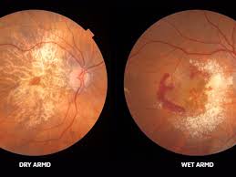
There are two types of ARMD: dry and wet.
Advanced ARMD is classified as either neovascular for wet ARMD or as atrophic or advanced dry ARMD.
Although wet form of ARMD accounts for only 10 to 15% of cases, it is responsible for the majority of cases of severe vision loss.
Dry ARMD is far more common.
Atrophic ARMD is characterized by atrophy of the retinal pigment epithelium and overlying neurosensory retina.
Wet ARMD is usually a more advanced disease state and is associated with rapid distortion and sudden loss of central vision.
The most vision impairing type of advanced age related macular degeneration is neovascular type,which is responsible for the majority of visual acuity loss in this disease.
The dry form of the disease involves the deposit of drusen under the retina and degeneration that affects approximately 90% of persons with age related maculate generation.
The leading cause of irreversible visual impairment in the elderly.
Age-related macular degeneration is classified as early and intermediate, with features of drusen and retinal pigment epithelium changes, and late AMD, which includes exudative AMD characterized by the presence of choroidal neovascularization.
Progression from early to intermediate, AMD is characterized by increasing drusen size and the appearance of pigmentary changes in the retina.
In AMD 25.9% of participants with larger drusen and pigmented abnormalities at baseline develop late stage AMD within five years.
There is migration of retinal pigment epithelium cells from their original attachment at the Bruch membrane into the more inner layers of the retina.
With confluence of areas of atrophy involving photoreceptors and retinal pigment epithelium, the process is known as geographic atrophy.
Abnormal growth of blood vessels in the macular region, neovascularization, is thought to be induced by increased expression of hypoxia driven vascular endothelial growth factor A which is released in response to stimuli, such as oxidative stress, and complement activation.
VEGF promotes angiogenesis by binding to its receptor and activating downstream pathways promoting endothelial cell proliferation.
Neovascular AMD causes visual changes when blood vessels leak, causing subretinal and intraretinal fluid accumulation, hemorrhages , and fibrosis-exudative neovascular AMD.
Neovascular age related macular degeneration is characterized by the abnormal growth of new blood vessels from choriocapillaries into the sub retinal pigment epithelial or sub retinal space, which can cause fluid in blood to leak.
Neovascular a or MD is characterized by proliferative neovascularization underneath the neurosensory retina.
AMD is characterized by focal or diffuse lipoprotein rich deposits called drusen, which form underneath the retinal epithelium, or by subretinal drusenoid deposits that accumulate under the neurosensory retina.
Drusen,the extracellular deposits, comprised of lipids, minerals, and proteins, and or implicated in the development and progression of AMD.
The observed angiogenesis is due to excessive vascular endothelial growth factor (VE GF).
Early AMD is associated with macular retinal yellowish deposits known as drusen, which are focal hypopigmentation or hyperpigmentation.
Progression from early to late stage AMD occurs at a rate of about 4% per year.
The Age-Related Eye Disease study showed a formulation of antioxidant vitamins and minerals could reduce the risk of progression from intermediate stage to late stage AMD by 25% over five years.
Exceeding recommended dietary allowances with antioxidant, vitamins and minerals can lead to adverse effects: kidney stones, fatigue, muscle weakness, decreased thyroid function, increased risk of hemorrhagic stroke, lung cancer, yellow skin, sideroblastic anemia, genitourinary symptoms, and upset stomach.
A degenerative disease of the central part of the retina, the macula, that results in a loss of central vision.
Central vision is essential for most daily activities.
ARMD characterized by a loss of visual acuity due to degeneration of the choriocapillaris, retinal pigment epithelium, and photoreceptors, usually beginning with drusen and pigmentary changes in Bruch’s membrane.
Affects 30 million to 50 million people worldwide.
The leading cause of irreversible blindness in developed countries in people aged 50 years and older.
More than 1.75 million persons in the United States were reported to have ARMD in 2000.
Prevalence increases exponentially every decade after age 50.
Prevalence in individuals older than 55 years is 1.6% and increases to about 13% in persons older than 84 years.
ARMD’s loss of central visual acuity leads to a reduction in activities of daily living, impaired mobility and an increased risk of falls, fractures, and depression.
It is associated with more than a doubling of all-cause mortality and more than a threefold higher risk of mortality not due to cardiovascular disease and cancer.
Age related macular degeneration may be a marker of frailty and aging or may be due to residual confounding factors indicative of aging.
Stages of ARMD: the early stage of ARMD is characterized by a macula with yellowish subretinal deposits referred to as drusen.
With early disease visual acuity may be stable for many years.
Loss of vision is a gradual process.
Patients with impaired dark adaptation are twice as likely to develop clinically evident age related macular degeneration and eight times as likely to advance beyond the earliest stages.
Advanced disease causes a significant loss of vision.
There are two types of ARMD, they are dry and wet types.
Dry ARMD is known as nonexudative, nonneovascular, or atrophic disease and is the most common form.
Dry ARMD occurs in about 90% of cases.
Loss of vision in dry ARMD is gradual and usually moderate.
In dry ARMD visual impairment includes fluctuating vision, difficulty reading, and limited vision at night or reduced illumination.
In dry ARMD the macula shows areas of depigmentation.
Wet ARMD is also referred to as exudative or neovascular ARMD
Wet ARMD ccounts for about 10% of cases.
Wet ARMD usually indicates a more advanced disease state.
Wet ARMD is associated with rapid vision distortion and sudden loss of central vision over a period of weeks to months.
In patients with wet ARMD, there is two times the expected prevalence of vitreomacular adhesion and are less likelihood to have a posterior vitreous detachment.
Fluid and exudate may accumulate underneath the retina in patients with wet ARMD causing macular edema, and if untreated, the neovascular membrane forms a macular scar and result in a sudden decrease in central vision.
Choroidal neovascularization refers to an advanced stage of wet ARMD that can lead to the development of choroidal vasculopathy.
The process progresses from drusen to the development of chorioneovascularization whereby the choriocapillaries cross Brucha’s membrane and spread laterally.
The etiology is multifactorial and involves genetic, environmental, metabolic, and functional factors.
Genes associated with AMD encode proteins in the complement pathway, lipid metabolism, DNA repair, collagen production, protein binding, and cell signaling.
Genetic factors are important in AMD with more than 30 gene polymorphisms associated with risk.
Genes strongly associated with the development and progression of AMD include: genes that include complement factor H as well as ARMS 2, and HTRA1.
Risk factors involved include aging, family history, smoking, high blood pressure, obesity, female gender, hypercholesterolemia, diabetes, obesity, sun and alcohol exposure, arteriosclerosis, and hypothyroidism.
Smoking, uncontrolled hypertension and BMI above 25, are linked to AMD.
Association between melatonin use and sowing AMD development.
The deterioration of the macula results in the loss of central vision, while peripheral vision remains intact, and patients with ARMD typically do not require canes or guide dogs.
Drusen are focal deposits of extracellular debris that typically form between the basal lamina of the retinal pigment epithelium and the inner collagenous layer of Bruch�s membrane.
Drusen are generally round and yellowish in color and are considered the hallmark of ARMD.
Drusen are classified as hard or soft, depending upon their borders.
Soft drusen are more commonly found in the macula and are associated with a higher risk development of age related macular degeneration.
Soft drusen are slightly larger than hard drusen and do not have well-defined margins.
Hard drusen tend to be smaller and are more well defined.
The features of the drusen may give an indication of the stage of ARMD.
Drusen contain lipids, carbohydrates, zinc, and at least 129 different proteins, including extracellular matrix.
In its early stages, ARMD is usually asymptomatic as the onset is gradual.
In some early cases there may be acute vision loss, blurred vision, scotomas, or chronic visual distortion.
About 13% of patients present with Charles Bonnet syndrome, in which mentally healthy patients experience loss of vision and complex visual hallucinations.
Charles Bonnet syndrome is benign and frequently regresses as the visual cortex adapts to the loss of vision.
Screening tests for ARMD include visual acuity tests, dilated funduscopic examination, optical coherence tomography, fluorescein angiography, indocyanine green angiography, fundus autofluorescence, and ultrasonography.
Optical coherence tomography imaging has enhanced ability to evaluate AMD lesions noninvasively and has largely replaced die based angiography in the management of most cases.
In advanced cases, referral to a retinal specialist may be required.
The Amsler grid is a 4×4-inch checkerboard chart that is an effective tool for monitoring the progression of ARMD at home.
Management is to identify and attack the disease in its early stages, slowing progression and vision loss.
Lifestyle changes and diet have been established as having a beneficial effect on preventing the disease and halting progression.
Thermal laser photocoagulation was the treatment of choice in the management of patients with wet ARMD.
The laser is directed toward the choroidal neovascularization to destroy it.
Laser photocoagulation is associated with a high rate of recurrence.
Various strategies including laser photocoagulation for neovascular ARMD, submacular surgery, external beam irradiation, proton beam irradiation, focal radiation, intravitreal steroids and transpupillary thermotherapy demonstrate no efficacy, or are associated with adverse effects.
The prognosis with neovascular ARMD has improved significantly with the verteporfin photodynamic therapy (PDT) and antiangiogenic therapy with intravitreal pegaptanib sodium, intravitreal bevacizumab, and intravitreal ranibizumab.
In photodynamic therapy (PDT) a combination of drugs and laser therapy.
In photodynamic therapy a verteporfin photosensitive compound is injected intravenously and is localized to the target site and then excited with laser light of a specific wavelength forming free radicals, which coagulate leaky subretinal vessels responsible for cellular injury in ARMD.
VEGF ihibition shows visual improvements of +6.9 letters to +11.3 letters can be achieved by using bevacizumab, ranibizumab, and aflibercept intravitreally in patients with choroidal neovascularization.
The most effective treatment of neovascular age related macular degeneration is intravitreal injection of anti-vascular endothelial growth factor (VEGF) antibodies.
Anti-– VEGF biological treatments, delivered by intravitreal injection are the first line therapy for treating and stabilizing exudative neovascular AMD as demonstrated by several randomized clinical trials.
Ranibizumab costs 40 times more than bevacizumab.
Age-related macular degeneration accounts for more than 50% of all blindness in United States.
Among individuals 75 years and older more than 25% have signs of age related maculopathy and 6-8% have advanced stages of disease.
Affects approximately 5% of U.S. population.
More than 8 million individuals have age-related macular degeneration in the U.S.
Prevalence twice that of Alzheimer’s disease.
Estimated 1.75 million people in the U.S. have advanced age-related macular degeneration, accounting for most cases of severe vision loss.
Is expected that the number of persons with advanced AMD will double over the next 20 years.
Patients with diabetes using glucagon-like peptide 1 receptor agonists (GLP-1 RAs) may face more than twice the risk for developing neovascular age-related macular degeneration compared with those not using the medications,
About 7.3 million people have early disease, which is usually associated with little or no vision loss, but increases the risk of developing advanced age related macular degeneration.
Age related disease associated with CFH gene polymorphisms, including Y402H on chromosome 1, and LOC387715 variant A69S on chromosome 10.
The advanced types of age related macular degeneration of geographic atrophy and neovascular disease associated with above polymorphisms.
Choroidal neovascularization, atrophy of retinal pigment epithelium associated with loss of central vision.
Hallmarks include the presence of hard or soft drusen and nonspecific pigment alterations at the retinal pigment epithelium.
First step in the diagnosis ARMD is clinical history followed by a comprehensive eye examination to include visual acuity, grid testing, and funduscopic evaluation after pharmacologic pupillary dilatation.
Both eyes should be examined by means of slit lamp bio microscopy to identify features of ARMD, classify the disease severity, and indicate the prognosis and treatment.
Color fundus photography represents a valuable tool for documenting whether in clinical trials are routine clinical settings, allows for effective comparisons during follow up visits.
Optical coherence tomography allows for three-dimensional visualization of ocular structures and detection retinal and subretinal fluid and to monitor exudative neovascular AMD treatment response.
Fundus autoflorescence imaging helps identify and monitor geographic atrophy progression.
Intravenous fluorescein angiography detects neovascularization reliably.
Geographic atrophy indicates advanced non-neovascular ARMD, whereas macular fluid or hemorrhage with underlying macular changes are consistent, indicating advanced neovascular ARMD.
Median age-related macular degeneration is defined as bilateral drusen.
Hallmark of the disease is the presence of drusen.
In most cases drusen are present bilaterally.
The presence of a few small hard drusen does not establish the diagnosis as these lesions are frequent over the age of 50 years and are a normal part of aging.
Excess drusen lead to damage of the retinal pigment.
Damage to the retinal pigment and chronic inflammation lead to retinal atrophy, geographic atrophy, secondary to the expression of antigenic cytokines.
Collagen or elastin abnormalities in Bruch’s membrane or the choroid may predispose to this process.
Early in the disease process vision may be normal and associated atrophic changes occur vision deteriorates.
Focal deposition of extracellular material, drusen, underneath retinal pigment epithelium.
Exudative, wet, age related macular degeneration is associated with abnormal neovascularization occurring under the retina.
Dry, nonexudative, age related macular degeneration is the most common type and represents 75-85% of cases.
Inflammation an important mechanism in this disease process with drusen shown to contain complement components immunoglobulins, regulators and anaphylatoxins.
Age related macular degeneration commonly non-neovascular, atrophic form associated with gradual vision impairment.
Neovascular type accounts for a relatively rapid severe loss of vision in about 10% of patients.
Age related-leading cause of vision loss of older patients.
Age related macular degeneration-associated with cigarette smoking and perhaps low levels of antioxidant nutrients.
Cigarette smoking is the most consistently reported environmental risk factor for AMD.
Age related-aspirin not beneficial in preventing process.
A combination of genetics and drinking instant coffee may increase a persons risk of developing dry AMD.
Melatonin use may decrease the prevalence and delay the progression of age related macular generation.
The Beaver Dam Eye Study assessed the use of ASA for 5 years and was not associated with Age-related macular degeneration, but use for 10 years associated with small but significant increase in risk of incident late and neovasvular AMD (Klein BEK et al).
In a study, which included more than 300,000 participants aged 55 or older, the use of low dose metformin was associated with a lower incidence of age-related macular degeneration (AMD).
The use of metformin was associated with a reduction in odds of developing AMD.
The association is dose-dependent, with low to moderate doses of metformin being associated with the greatest benefit.
When assessing doses greater than 1080 g, there was no association with reduced odds of developing AMD at 2 years.
Among patients with diabetes, metformin use was associated with a decreased risk of AMD in patients without existing diabetic retinopathy, but was considered a risk factor for patients with existing diabetic retinopathy.
Metformin use over 2 years in adults aged 55 years and older is associated with 5% to 10% reduced odds ratio of developing AMD.
A dose-dependent association of this potential protective effect is present, with low to moderate doses of metformin being associated with the lowest odds ratio for the development of AMD.
Age related-develops slowly as a result of accumulated damage over several years.
Nutritional supplements, including vitamins, carotenoids, and trace elements such as zinc may be associated with slower rates of AMD progression.
Consuming recommended fish twice a week, and daily amounts of vegetables are associated with lower rates of AMD risk.
AMD affects central vision and complete blindness typically does not occur.
AMD can cause significant visual impairment and legal blindness, making individuals ineligible to drive and affect capacity to read and recognize faces.
Age related-atrophy of choriocapillaris with decreased number and diameter of capillaries in the macular region of the eye.
Inverse relationship with omega-3 fatty acid intake and increased intake of linoleic acid associated with a higher prevalence of macular degeneration.
Higher intake of vegetable, monounsaturated and polyunsaturated fats are associated with higher risk of macular degeneration.
Observational studies suggested higher dietary intake of lutein plus zeaxanthin and eicosapentaenoic acid are associated with these decreased risk of developing age-related macular degeneration.
Lutein and zeaxanthin are the major components of macular pigment.
Omega-3 long chain polyunsaturated fatty acid docosahexaenoic acid (DHA) is a major component of the retina
Direct association between homocysteine concentration in the blood and risk of disease.
Elevated CRP level is an independent risk factor for age-related macular degeneration.
The Age-Related Eye DiseaseStudy (AREDS) indicated that taking a high formulation of antioxidants and zinc significantly reduces the risk of advanced disease and its associated vision loss for persons at intermediate disease stages.
The micronutrients used in the above study include vitamin C, zinc, vitamin E, copper and beta carotene. (Subsequently removing beta carotene for safety reasons).
In the above study there was reduced risk of developing advanced AMD by 25% at five years.
Because the rate of developing late stage AMD among patients with early AMD is low at 1.3% after five years, supplementation with antioxidant vitamins and minerals is recommended for patients who have already progressed to intermediate or late stage AMD in at least one eye.
Patients should have lifestyle intervention with counseling against smoking, and management of hypertension.
Treatment for wet age related macular degeneration include laser surgery, photodynamic therapy, and injections into the eye of anti-angiogenic medications.
Intraocular drugs that inhibit vascular endothelial growth factor are available for the treatment of neovascular age-related macular degeneration and there is presently no effective therapy for atrophic AMD.
Vascular endothelial growth factor targeting therapies can prevent vision loss and have revolutionized care in patients with neovascular AMD.
Intravitreal injection of anti-VEGF agents to treat choroidal neovascularization reduces the risk of moderate vision loss, and visual acuity to less than 10% over a 24 month period, as compared with approximately 50% in the control group.
On average patients receive seven or eight injections during initial 12 month period of therapy.
Bevacizumab (Avastin) and ranibizumab (Lucentis) have equivalent results on visual acuity when treated with intravitreal injections (CATT Research Group).
Use of Bevacizumab and ranibizumab used on a as needed basis had results equivalent to monthly scheduled treatments (CATT Research Group).
Bevacizumab most common drug treatment for AMD.
Ranibuzumab costs 40 times as much as Bevacizumab.
The goal of treatment with anti-VEGF agents is resolution of leakage of blood vessels in the macula and hemorrhage associated with neovascular AMD.
Delays in the treatment of exudative neovascular AMD is associated with visual loss.
Treatment for exudative neovascular AMD should commence within 14 days of the initial diagnosis.
Approximately half of patients followed for 5 to 7 years retain at least 20/40 vision.
A review of 16 randomized clinical trials concluded that serious adverse ocular events after intravitreal rejection of anti-VEGF agents, such as endophthalmitis, retinal detachment, traumatic lens injury, increased intraocular pressure are infrequent.
In patients with inadequate response to pharmacotherapy photodynamic therapy may be beneficial.
Levodopa treatments improve visual outcomes and stabilize neovascular age related macular degeneration related retinal changes.
Levodopa reduces VEGF levels and may be effective as an adjuvant anti-VEGF injection therapy.
Faricimab is the first bispecific monoclonal antibody to target both vascular endothelial growth factor (VEGF)[1] and angiopoietin 2 (Ang-2).
Faricimab targeting these pathways stabilizes blood vessels in the retina.
Anti-VEGF treatments do not prevent atrophy and even with aggressive treatment many patients with subretinal neovascularization eventually lose vision.
Vision loss from geographic atrophy can also occur in the absence of neovascularization, when photo receptors, RPE cells, and the choricapillaris capillaries vessels that supply blood to the outer retina degenerate.
Geographic atrophy causes loss of cells that respond to light a prosthetic system called PRIMA (photovoltaic retina implant microwave) can restore light perception to the central retina.
The PRIMA system includes a photovoltaic device, surgically implanted beneath the central retina, that receives in the year infrared red light projected from a camera on a pair of glasses.
The photovoltaic pixels in the device convert infrared light from the camera into electrical impulses that stimulate the retinal, bipolar cells, which connect with retinal ganglion cells and send visual information through the optic nerves to the brain for visual perception.
The device compensates for the missing macular vision cells by sending information through the inner retinal and optic nerve pathways to the brain.
One reply on “Macular degeneration (AMD)”
[…] See ((Macular degeneration)). […]