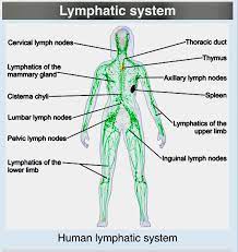
 The lymphatic system is an extensive network of lymphatic blood vessels and lymph nodes, with lymphoid tissue in the spleen, gastrointestinal tract, tonsils and adenoids.
The lymphatic system, or lymphoid system, is an organ system that is part of the immune system.
It is complementary to the circulatory system.
Clearance of excess fluid and interstitial solutes is critical for tissue homeostasis.
In the peripheral tissues soluble material, proteins and fluid from the interstitial space are returned to the general circulation by the lymphatic system.
In the peripheral organs, the lymphatic system performs important immune functions and runs parallel to the blood circulatory system to provide a secondary circulation that transports excess interstitial fluid, proteins, and metabolic waste products from the systemic tissues back into the blood.
The lymphatic system acts like an overflow and can return a lot of excess fluid back to the bloodstream.
Lymph is a clear fluid that is carried by the lymphatic vessels back to the heart for re-circulation.
The lymphatic system is open, unlike the circulatory system that is a closed system
The lymphatic network extends throughout all parts of the peripheral tissues and the density of lymph vessels correlates with the rate of tissue metabolism.
The lymphatic system can be overwhelmed, and if there is simply too much fluid, or if the lymphatic system is congested, causing swellings in legs, ankles, feet, abdomen or any other part of the body.
The human circulatory system processes an average of 20 litres of blood per day through capillary filtration, which removes plasma from the blood.
Roughly 17 litres of the filtered blood is reabsorbed directly into the blood vessels, while the remaining three litres are left in the interstitial fluid.
One of the main functions of the lymphatic system is to provide an accessory return route to the blood for the surplus three litres.
Interstitial fluid is recirculated by the lymphatics, which are tributaries to the arterial venous circulation that start as opened vessels in the interstitium.
The importance of lymphatic flow is especially evident when the lymphatic system becomes obstructed.
In lymphatic associated diseases, such as elephantiasis, the impact of such an obstruction may be dramatic.
The efficient removal of soluble proteins from the interstitial fluid is critical to the regulation of both colloidal osmotic pressure and homeostatic regulation of the fluid volume of the body.
Lymph vessels transport lymph fluid with protein, immune cells and waste products to lymph nodes and spleen where substances and cells are filtered.
The efficient removal of soluble proteins from the interstitial fluid is critical to the regulation of both colloidal osmotic pressure and homeostatic regulation of the fluid volume of the body.
Lymphatics have valves for one-way flow down the shallow pressure gradient to the thoracic duct, which returns to the subclavian vein.
Lymph flow is screened for pathogenic agents through lymph nodes.
Food, stuffs, nutrients, and micro nutrients, such as calcium, magnesium, zinc, iron, and copper, or absorbed in the gut interstitium of the villus, which contains a central intestinal, lymphatic or lacteal.
Other blood constituents, including hematopoietic cells, fats, and vitamins can also enter the lymph.
Lymph is ultimately returned to the circulation via the right lymphatic duct and the thoracic duct.
Adult lymphatic vasculature is quiescent except when tissues or organs are undergoing repair or regeneration and during pathologic processes such as tumor growth and metastases.
Lymphangiogenic factors include vascular endothelial growth factor, fibroblast growth factor, angiopoietin, platelet derived growth factor and hepatocyte growth factor.
Epithelial cells connected by tight junctions guard the integrity and composition of the gut interstitium.
Proteins losing enteropathy occurs when the intestinal epithelium is breached and the intestitial fluid leaks into the gut lumen.
Another function of lymph is that of immune defense.
Lymph similar to plasma, and contains waste products and cellular debris, together with bacteria and proteins.
The cells of the lymph are mostly lymphocytes.
Associated lymphoid organs are composed of lymphoid tissue, and are the sites either of lymphocyte production or of lymphocyte activation.
Lymphoid organs include the lymph nodes, where the highest lymphocyte concentration is found, the spleen, the thymus, and the tonsils.
Lymphocytes are initially generated in the bone marrow.
The lymphoid organs also contain other types of cells-stromal cells for support.
Lymphoid tissue is also associated with mucosas such as mucosa-associated lymphoid tissue (MALT).
Circulating blood leaks into the surrounding tissues of the body by capillary action, carrying nutrients to the cells.
The fluid bathes the tissues as interstitial fluid.
The interstitial fluid collects waste products, bacteria, and damaged cells, and then drains as lymph into the lymphatic vessels.
Lymphatic vessels carry the lymph throughout the body, passing through numerous lymph nodes that filters out unwanted materials such as bacteria and damaged cells.
Lymph then passes into much larger lymph vessels known as lymph ducts.
The right lymphatic duct drains the right side of the region and the much larger left lymphatic duct, known as the thoracic duct, drains the left side of the body.
The lymphatic system carries excess plasma filtered from the capillaries as interstitial fluid between cells, away from the body tissues in an accessory route to return the excess fluid back to the blood circulation as lymph.
The passage of lymph takes much longer than that of blood.
The lymphatic system is essential for the functioning of the blood circulatory system, for without it the blood would become depleted of fluid.
The lymphatic system works together with the immune system.
The lymphatic ducts empty into the subclavian veins to return to the blood circulation.
Lymph is moved through the system by muscle contractions.
The lymphatic ducts empty into the subclavian veins to return to the blood circulation.
Lymph is moved through the system by muscle contractions.
The thymus and the bone marrow constitute the primary lymphoid organs involved in the production and early clonal selection of lymphocyte tissues.
Bone marrow is responsible the creation of T cell precursors and the production and maturation of B cells.
From the bone marrow, B cells immediately join the circulatory system and travel to secondary lymphoid organs in search of pathogens.
T cells, on the other hand, travel from the bone marrow to the thymus, where they develop further and mature.
Mature T cells then join B cells in search of pathogens.
The other 95% of T cells begin a process of apoptosis, a form of programmed cell death.
The thymus increases in size from birth in response to postnatal antigen stimulation.
It is most active during the neonatal and pre-adolescent periods.
The thymus is located between the inferior neck and the superior thorax.
At puberty, by the early teens, the thymus begins to atrophy and regress, with adipose tissue mostly replacing the thymic stroma.
However, residual T cell lymphopoiesis continues throughout adult life, providing some immune response.
The thymus is where the T lymphocytes mature and become immunocompetent.
The loss/lack of the thymus results in severe immunodeficiency and subsequent high susceptibility to infection.
The thymus consists of epithelium.
T cells mature from thymocytes, proliferate, and undergo a selection process in the thymic cortex before entering the medulla to interact with epithelial cells.
The thymus provides an inductive environment for the development of T cells from hematopoietic progenitor cells.
Thymic stromal cells allow for the selection of a functional and self-tolerant T cells.
The secondary lymphoid organs include lymph nodes and the spleen.
They maintain mature naive lymphocytes and initiate an adaptive immune response.
The secondary lymphoid organs are the sites where lymphocytes are activated by antigens,leading to clonal expansion, and affinity maturation.
Mature lymphocytes recirculate between the blood and the secondary lymphoid organs until they encounter their specific antigen.
The main functions of the spleen are:
to produce immune cells to fight antigens
to remove particulate matter and aged blood cells, mainly red blood cells
to produce blood cells during fetal life.
The spleen synthesizes antibodies in its white pulp.
The spleen removes antibody-coated bacteria and antibody-coated blood cells by way of blood and lymph node circulation.
The white pulp of the spleen provides immune function due to the lymphocytes that are housed there.
The spleen’s red pulp which is responsible for getting rid of aged red blood cells, as well as pathogens.
This is carried out by macrophages present in the red pulp.
The spleen contains, in its reserve, half of the body’s monocytes within the red pulp, which upon moving to injured tissue turn into dendritic cells and macrophages while promoting tissue healing.
The spleen is a center of activity of the mononuclear phagocyte system.
The spleen is analogous to a large lymph node, as its absence causes a predisposition to certain infections.
The spleen also is responsible for recycling some RBC components and discarding others.
Hemoglobin is broken down in the spleen into amino acids that are reused.
Like the thymus, the spleen has only efferent lymphatic vessels.
Both the short gastric arteries and the splenic artery supply it with blood.
The germinal centers are supplied by arterioles called penicilliary radicles.
Until the fifth month of prenatal development, the spleen creates red blood cells; after birth, the bone marrow is solely responsible for hematopoiesis.
Lymphatic tissues begin to develop by the end of the fifth week of embryonic development.
Lymphatic vessels develop from lymph sacs that arise from developing veins, which are derived from mesoderm.
Lymph sacs appear at jugular lymph sacs at the internal jugular and subclavian veins.
From the jugular lymph sacs, lymphatic capillary plexuses spread to the thorax, upper limbs, neck, and head.
Each jugular lymph sac retains at least one connection with its jugular vein, the left one developing into the superior portion of the thoracic duct.
The spleen develops from mesenchymal cells between layers of the dorsal mesentery of the stomach.
The thymus arises as an outgrowth of the third pharyngeal pouch.
The lymphatic system is responsible for the removal of interstitial fluid from tissues.
The lymphatic system absorbs and transports fatty acids and fats as chyle from the digestive system.
It transports white blood cells to and from the lymph nodes into the bones.
The lymph transports antigen-presenting cells, such as dendritic cells, to the lymph nodes where an immune response is stimulated.
Nutrients in food are absorbed via intestinal vili to blood and lymph.
Long-chain fatty acids-and other lipids with similar fat solubility are absorbed to the lymph and move in it enveloped inside chylomicrons.
They move via the thoracic duct of the lymphatic system and finally enter the blood via the left subclavian vein, thus bypassing the liver’s first-pass metabolism completely.
Lymph vessels (lacteals) are at the beginning of the gastrointestinal tract, predominantly in the small intestine.
Most nutrients absorbed by the small intestine are passed on to the portal venous system to drain via the portal vein into the liver for processing.
Lipids are passed on to the lymphatic system to be transported to the blood circulation via the thoracic duct.
However, medium-chain triglycerides are fatty acid esters of glycerol that passively diffuse from the GI tract to the portal system.
The enriched lymph originating in the lymphatics of the small intestine is called chyle.
The nutrients that are released into the circulatory system are processed by the liver, having passed through the systemic circulation.
The lymphatic system’s role in the body’s immune system, is as the primary site for cells relating to adaptive immune system including T-cells and B-cells.
Cells in the lymphatic system react to antigens presented or found by the cells directly or by other dendritic cells.
With antigen recognition an immunological cascade begins involving the activation and recruitment of more and more cells, the production of antibodies and cytokines and the recruitment of other immunological cells such as macrophages.
Lymphatic drainage of various organs is important in the diagnosis, prognosis, and treatment of cancer, as the lymphatic system is responsible for carrying cancerous cells between the various parts of the body (metastasis).
Intervening lymph nodes can trap the cancer cells.
If lymph nodes are not successful in destroying the cancer cells the nodes may become sites of secondary tumours.
Lymphadenopathy refers to one or more enlarged lymph nodes, which are generally reactive in response to infection or inflammation.
Generalized lymphadenopathy may be caused by infections such as infectious mononucleosis, tuberculosis and HIV, connective tissue diseases such as SLE and rheumatoid arthritis, and cancers, including both cancers of tissue within lymph nodes, and metastasis of cancerous cells from other parts of the body, that have arrived via the lymphatic system.
Lymphedema is the swelling caused by the accumulation of lymph.
Lymphedema may occur if the lymphatic system is damaged or has malformations.
Lymphedema usually affects limbs, though the face, neck and abdomen may also be affected.
In an extreme state, elephantiasis, the edema progresses to the extent that the skin becomes thick with an appearance similar to the skin on elephant limbs.
Elephantiasis causes are unknown in most cases, but sometimes an infection, usually caused by a parasitic disease, such as lymphatic filariasis.
Lymphangiomatosis is a disease involving multiple cysts or lesions formed from lymphatic vessels.
Lymphedema can also occur after surgical removal of lymph nodes in the axilla or groin.
Cancer of the lymphatic system can be primary or secondary: Lymphoma refers to cancer that arises from lymphatic tissue.
Lymphoid leukemias and lymphomas are now considered to be tumors of the same type of cell lineage.
They are called leukemia when in the blood or marrow and lymphoma when in lymphatic tissue.
Lymphoma is generally considered as either Hodgkin lymphoma or non-Hodgkin lymphoma.
Hodgkin lymphoma is characterised by a particular type of cell, called a Reed–Sternberg cell, visible under microscope. It is associated with past infection with the Epstein–Barr virus, and generally causes a painless rubbery lymphadenopathy. It is staged, using Ann Arbor staging.
Chemotherapy generally involves the ABVD and may also involve radiotherapy.
Non-Hodgkin lymphoma is a cancer characterized by increased proliferation of B-cells or T-cells, generally occurs in an older age group than Hodgkin lymphoma.
It is treated according to whether it is high-grade or low-grade, and carries a poorer prognosis than Hodgkin lymphoma.
Lymphangiosarcoma is a malignant soft tissue tumour, whereas lymphangioma is a benign tumour occurring frequently in association with Turner syndrome.
Lymphangioleiomyomatosis is a benign tumour of the smooth muscles of the lymphatics that occurs in the lungs.
Other processes involving the lymphatic system include:
Castleman’s disease
Chylothorax
Kawasaki disease
Kikuchi disease
Lipedema
Lymphangitis
Lymphatic filariasis
Lymphocytic choriomeningitis
Solitary lymphatic nodule