 Suspensory muscle of the duodenum connects the duodenum of the small intestine to the diaphragm.
Suspensory muscle of duodenum, is also known as the ligament of Treitz.
The suspensory muscle of the duodenum attaches to the duodenojejunal flexure, behind the pancreas.
Arises from the connective tissue around the celiac trunk and superior mesentery artery and inserts into the third and fourth portions of the duodenum and frequently into the duodenal-jejunal flexure, as well.
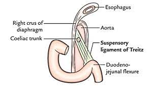
The suspensory muscle arises from the right crus of the diaphragm as it passes around the esophagus, continues as connective tissue around the stems of the celiac trunk (celiac artery) and superior mesenteric artery, passes behind the pancreas, and enters the upper part of the mesentery, inserting into the junction between the duodenum and jejunum, the duodenojejunal flexure.
The suspensory muscle of duodenum is a thin muscle connecting the junction between the duodenum, jejunum, and duodenojejunal flexure to connective tissue surrounding the superior mesenteric artery and celiac artery.
The suspensory muscle of the duodenum facilitates movement of food.
Its embryological role in fixating jejunum during gut rotation.
It marks the formal division between the first and second parts of the small intestine, the duodenum and the jejunum.
When it contracts it opens the duodenojejunal junction and permits the flow of chyme.
While referred to as a ligament it is a suspensory muscle and ligament due to its composition and function.
Important landmark when looking for the presence of malrotation of the gut, as it is abnormally located when this process is present in children.
marks the difference between the upper and lower gastrointestinal tracts, which is relevant in clinical medicine as it may determine the source of bleeding in the gastrointestinal tract.
The suspensory muscle is derived from mesoderm and plays a role in the embryological rotation of the gut, by offering a point of fixation for the rotating gut.
It helps digestion by widening the angle of the duodenojejunal flexure.
The superior mesenteric artery syndrome is a rare abnormality caused by a congenitally short suspensory muscle.
The duodenum and the jejunum are the first and second parts of the small intestine, respectively, and the suspensory muscle of the duodenum marks their formal division.
Anatomic variation exists, in terms of length and point of attachment of the sensory suspensory muscle of the duodenum.
The muscle only solely attaches to the duodenojejunal flexure in about 8% of people, and 40 to 60% of the time attaches additionally to the third and fourth parts of the duodenum; and 20 to 30% of the time it only attaches to the third and fourth parts.
Separate multiple attachments of this muscle not that uncommon.
The muscle may be divided into two sections: a ligamentous portion attaching the right crus of diaphragm to the connective tissue surrounding the celiac artery and superior mesenteric artery; and a lower muscular portion from the connective tissue attaching to the duodenum.
With contraction the sensory muscle of the duodenum, by virtue of connections to the third and fourth parts of the duodenum, widens the angle of the duodenojejunal flexure, allowing movement of the intestinal contents.
It is an important landmark to note bowel malrotation of the gut, a syndrome often suspected in young children when they have episodes of recurrent vomiting.
A normal location of the ligament of Treitz by radiological imaging is critical in ruling out malrotation of the gut in a child, as it is abnormally located when malrotation is present.
During a Whipple’s procedure, the ligament of Treitz is separated from the duodenum and preserved.
When the remaining jejunum is anastamosed with the pylorus of the stomach, it may be passed through the ligament.
Superior mesenteric artery syndrome (SMA) is an extremely rare life-threatening condition that can either be congenital and chronic, or induced and acute, and it is characterized by compression of the duodenum between the abdominal aorta and the superior mesenteric artery, and may, when congenital, result from a short suspensory muscle.