
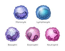
White blood cells (leukocytes), also called immune cells or immunocytes, are cells of the immune system that are involved in protecting the body against both infectious disease and foreign entities.
White blood cells are generally larger than red blood cells.
Leukocytes include three main subtypes: granulocytes, lymphocytes and monocytes.
All white blood cells are produced and derived from multipotent cells in the bone marrow known as hematopoietic stem cells.
Leukocytes are found throughout the body, including the blood and lymphatic system.
All white blood cells have nuclei, which distinguishes them from the other blood cells, the anucleated red blood cells (RBCs) and platelets.
The different white blood cells are usually classified by their cell lineage-myeloid cells or lymphoid cells.
White blood cells, part of the body’s immune system as they help the body fight infection and other diseases.
Types of white blood cells are granulocytes (neutrophils, eosinophils, and basophils), and agranulocytes (monocytes, and lymphocytes (T cells and B cells)).
Myeloid cells (myelocytes) include neutrophils, eosinophils, mast cells, basophils, and monocytes.
Monocytes are further subdivided into dendritic cells and macrophages.
Monocytes, macrophages, and neutrophils are phagocytic.
Lymphoid cells (lymphocytes) include T cells (subdivided into helper T cells, memory T cells, cytotoxic T cells), B cells (subdivided into plasma cells and memory B cells), and natural killer cells.
A key function of leukocytes is their ability to migrate and extravasation into tissues, facilitated by cell surface molecules that promote adhesion to the vascular endothelium.
The beta to integrated molecule, CD 18, is the common component of four molecular complexes that play a crucial role in leukocyte trafficking, phagocytosis, cytokinine release, and signaling in cells of innate and adaptive immune systems.
An excess of white blood cells is usually due to infection or inflammation.
A high white blood cell count could indicate certain blood cancers or bone marrow disorders.
The normal white cell count is usually between 4 billion/L and 11 billion/L: This is usually expressed as 4,000 to 11,000 white blood cells per microliter of blood.
White blood cells make up approximately 1% of the total blood volume in a healthy adult, making them substantially less numerous than the red blood cells at 40% to 45%.
The WBC differential is usually reported as part of a complete blood count (CBC). Results are provided as both percentages and absolute numbers for each cell type, and compared to reference ranges typical for adults.
This test may be performed by automated machines or by manual counting under a microscope, especially when abnormal results are suspected.
Key reference ranges for adults (as a percentage of total WBCs): • Neutrophils: 50–70% • Lymphocytes: 18–42% • Monocytes: 2–11% • Eosinophils: 1–3% • Basophils: 0–2%
An increase in the number of leukocytes over the upper limits is called leukocytosis.
A decrease below the lower limit is called leukopenia, which indicates a weakened immune system.
White cells are found in the buffy coat, a thin, typically white layer of nucleated cells between the sedimented red blood cells and the blood plasma.
The buffy coat may sometimes be green if there are large amounts of neutrophils in the sample, due to the heme-containing enzyme myeloperoxidase that they produce.
All white blood cells are nucleated, which distinguishes them from the anucleated red blood cells and platelets.
Types of leukocytes can be classified:
Two pairs of broadest categories classify them either by structure (granulocytes or agranulocytes) or by cell lineage (myeloid cells or lymphoid cells).
These categories can be further divided into the five main types: neutrophils, eosinophils, basophils, lymphocytes, and monocytes.
A good way to remember the relative proportions of WBCs is Never Let Monkeys Eat Bananas.
Monocytes and neutrophils are phagocytic.
Granulocytes are distinguished from agranulocytes by their nucleus shape, lobed versus round, that is, polymorphonuclear versus mononuclear, and by their cytoplasm granules either present or absent.
Myeloid cells (neutrophils, monocytes, eosinophils and basophils) are distinguished from lymphoid cells (lymphocytes) by hematopoietic lineage or cellular differentiation lineage.
Lymphocytes can be further classified as T cells, B cells, and natural killer cells.
Neutrophils are the most abundant white blood cell, constituting 60–70% of the circulating leukocytes.
Neutrophils defend against bacterial or fungal infection.
They are usually first responders to microbial infection; activity and death in large numbers form pus.
They are commonly referred to as polymorphonuclear (PMN) leukocytes.
They have a multi-lobed nucleus, which consists of three to five lobes connected by slender strand, giving the neutrophils the appearance of having multiple nuclei, hence the name polymorphonuclear leukocyte.
The cytoplasm may look transparent because of fine granules that are pale lilac when stained.
Neutrophils are active in phagocytosing bacteria and are present in large amount in the pus of wounds.
Neutrophils are not able to renew their lysosomes that are used in digesting microbes and die after having phagocytosed a few pathogens.
Neutrophils are the most common cell type seen in the early stages of acute inflammation.
The average lifespan of inactivated human neutrophils in the circulation has been reported by different approaches to be between 5 and 135 hours.
Eosinophils compose about 2–4% of white blood cells in circulating blood.
The eosinophil count fluctuates throughout the day, seasonally, and during menstruation.
It rises in response to allergies, parasitic infections, collagen diseases, and disease of the spleen and central nervous system.
They are rare in the blood, but numerous in the mucous membranes of the respiratory, digestive, and lower urinary tracts.
Eosinophils primarily deal with parasitic infections, and are also the predominant inflammatory cells in allergic reactions.
The most important causes of eosinophilia: include allergies such as asthma, hay fever, and hives; and parasitic infections.
Eosinophils secrete chemicals that destroy large parasites, such as hookworms and tapeworms, that are too big for any one white blood cell to phagocytize.
In general, their nuclei are bi-lobed, and lobes are connected by a thin strand.
The cytoplasm is full of granules that assume a characteristic pink-orange color with eosin staining.
Basophils are chiefly responsible for allergic and antigen response by releasing the chemical histamine causing the dilation of blood vessels.
Because they are the rarest of the white blood cells (less than 0.5% of the total count) and share physicochemical properties with other blood cells.
They can be recognized by several coarse, dark violet granules, giving them a blue hue.
The nucleus is bi- or tri-lobed, but it is hard to see because of the number of coarse granules that hide it.
They secrete two chemicals that aid in the body’s defenses: histamine and heparin.
Histamine is responsible for widening blood vessels and increasing the flow of blood to injured tissue, and makes blood vessels more permeable so neutrophils and clotting proteins can get into connective tissue more easily.
Heparin is an anticoagulant that inhibits blood clotting and promotes the movement of white blood cells into an area.
Basophils can also release chemical signals that attract eosinophils and neutrophils to an infection site.
Lymphocytes are much more common in the lymphatic system than in blood.
Lymphocytes are distinguished by having a deeply staining nucleus that may be eccentric in location, and a relatively small amount of cytoplasm.
Lymphocyte B cells make antibodies that can bind to pathogens, block pathogen invasion, activate the complement system, and enhance pathogen destruction.
CD4+ T helper lymphocytes cells: T cells displaying co-receptor CD4 are known as CD4+ T cells.
These cells have T-cell receptors and CD4 molecules that, in combination, bind antigenic peptides presented on major histocompatibility complex (MHC) class II molecules on antigen-presenting cells.
Helper T cells make cytokines and perform other functions that help coordinate the immune response.
In HIV infection, these T cells are the main index to identify the individual’s immune system integrity.
CD8+ cytotoxic T cells displaying co-receptor CD8 are known as CD8+ T cells.
These cells bind antigens presented on MHC I complex of virus-infected or tumor cells and kill them.
Natural killer cells are able to kill cells of the body that do not display MHC class I molecules, or display stress markers such as MHC class I polypeptide–related sequence A (MIC-A).
Decreased expression of MHC class I and up-regulation of MIC-A can happen when cells are infected by a virus or become cancerous.
Monocytes, the largest type of white blood cell, share the phagocytosis function of neutrophils, but are much longer lived.
Monocytes present pieces of pathogens to T cells so that the pathogens may be recognized again and killed.
This causes an antibody response to be mounted.
Monocytes eventually leave the bloodstream and become tissue macrophages.
Tissue macrophages remove dead cell debris as well as attack microorganisms.
Neither dead cell debris nor attacking microorganisms can be dealt with effectively by the neutrophils.
Unlike neutrophils, monocytes are able to replace their lysosomal contents and are thought to have a much longer active life.
Monocytes have a kidney-shaped nucleus, are typically not granulated, and also possess abundant cytoplasm.
Some leucocytes migrate into the tissues of the body to take up a permanent residence at that location rather than remaining in the blood.
Fixed macrophages in the liver, which become known as Kupffer cells, serving a role in the immune system.
The categories of white blood cell disorders divide them quantitatively into those causing excessive numbers or proliferative disorders, and those causing insufficient numbers, leukopenias).
Leukocytosis is usually healthy (e.g., fighting an infection), but it also may be dysfunctionally proliferative.
Proliferative disorders of white blood cells can be classed as myeloproliferative or lymphoproliferative, autoimmune, or neoplastic.
White blood cell disorders include those in which the blood cells do not function normally.
Neoplasia of white blood cells can be benign but is often malignant, classified as leukemias and lymphomas
Leukopenias consist of a range of disorders that can cause decreases in white blood cells.
This type of white blood cell decreased is usually the neutrophil, referred to as neutropenia or granulocytopenia.
A decrease in lymphocytes is called lymphocytopenia or lymphopenia.
Neutropenia can be acquired or intrinsic.
A decrease in levels of neutrophils is due to either decreased production of neutrophils or increased removal from the blood.
Medications associated with neutropenia-chemotherapy, sulfas or other antibiotics, phenothiazines, benzodiazepines, antithyroid medications, anticonvulsants, quinine, quinidine, indometacin, procainamide, thiazides Radiation
Toxins associated with neutropenia alcohol, benzenes
Intrinsic disorders associated with neutropenia – Fanconi’s, Kostmann’s, cyclic neutropenia, Chédiak–Higashi
Immune dysfunction associated with neutropenia-connective tissue diseases, AIDS, rheumatoid arthritis.
Blood cell dysfunctions associated with neutropenia– megaloblastic anemia, myelodysplasia,Bone marrow failure, Bone marrow transplant, acute leukemia
Any major infection, starvation, hypersplenism can be associated with neutropenia
Lymphocytopeni-defined as total lymphocyte count below 1.0 billion/L.
The cells most commonly affected are CD4+ T cells.
Like neutropenia, lymphocytopenia may be acquired or intrinsic.
Causes of lymphopenia:
Inherited immune deficiency – severe combined immunodeficiency, common variable immunodeficiency, ataxia–telangiectasia, Wiskott–Aldrich syndrome, immunodeficiency with short-limbed dwarfism, immunodeficiency with thymoma, purine nucleoside phosphorylase deficiency, genetic polymorphism Blood cell dysfunction – aplastic anemia Infectious diseases – viral (AIDS, SARS, West Nile encephalitis, hepatitis, herpes, measles, others), bacterial (TB, typhoid, pneumonia, rickettsiosis, ehrlichiosis, sepsis), parasitic (acute phase of malaria) Medications – chemotherapy (antilymphocyte globulin therapy, alemtuzumab, glucocorticoids) Radiation Major surgery Miscellaneous – ECMO, kidney or bone marrow transplant, hemodialysis, kidney failure, severe burns, celiac disease, severe acute pancreatitis, sarcoidosis, protein-losing enteropathy, strenuous exercise, carcinoma Immune dysfunction – arthritis, systemic lupus erythematosus, Sjögren syndrome, myasthenia gravis, systemic vasculitis, Behçet’s-like syndrome, dermatomyositis, granulomatosis with polyangiitis Nutritional/Dietary – alcohol use disorder, zinc deficiency
Like neutropenia, symptoms and treatment of lymphocytopenia are directed at the underlying cause of the change in cell counts.
Leukocytosis
An increase in the number of white blood cells in circulation is called leukocytosis.
This increase is most commonly caused by inflammation.
There are four major causes: increased production in bone marrow, increased release from storage in bone marrow, decreased attachment to veins and arteries, decreased uptake by tissues.
Leukocytosis may affect one or more cell lines and can be neutrophilic, eosinophilic, basophilic, monocytosis, or lymphocytosis.
Neutrophilia is an increase in the absolute neutrophil count in the peripheral circulation.
Normal blood values vary by age.
Neutrophilia can be caused by a direct problem with blood cells with primary disease It can also occur as a consequence of an underlying disease as a secondary process.
Most cases of neutrophilia are secondary to inflammation.
Conditions with normally functioning neutrophils – hereditary neutrophilia, chronic idiopathic neutrophilia.
Pelger–Huët anomaly
Down syndrome
Leukocyte adhesion deficiency
Familial cold urticaria
Leukemia-CML and other myeloproliferative disorders
Surgical removal of spleen
Secondary causes
Infection
Chronic inflammation – especially juvenile idiopathic arthritis, rheumatoid arthritis, Still’s disease, Crohn’s disease, ulcerative colitis, granulomatous infections and chronic hepatitis
Cigarette smoking – occurs in 25–50% of chronic smokers and can last up to 5 years after quitting
Stress – exercise, surgery, general stress
Medication induced – corticosteroids, β-agonists, lithium
Cancer – by growth factors secreted by the tumor or invasion of bone marrow by the cancer
Increased destruction of cells in peripheral circulation can stimulate bone marrow and can occur in hemolytic anemia and idiopathic thrombocytopenic purpura.
Eosinophilia
A normal eosinophil count is considered to be less than 0.65×10⁹/L.
Eosinophil counts are higher in newborns and vary with age, time being lower in the morning and higher at night, exercise, environment, and exposure to allergens.
Eosinophilia is never a normal lab finding and efforts should always be made to discover the underlying cause.