
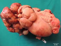
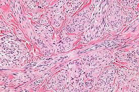
 Leiomyosarcoma is a malignant smooth muscle tumor requiring multimodal treatment with surgery as the cornerstone for localized disease and systemic therapy for advanced/metastatic disease.
Management is stratified by anatomic location (uterine vs. non-uterine), stage, and resectability. For resectable localized leiomyosarcoma, the primary treatment is surgical resection with oncologically appropriate margins (R0 resection).
Multimodality treatment is critical, with neoadjuvant or adjuvant radiation therapy recommended for most Stage II-III extremity and body wall tumors to improve local control.
The choice between neoadjuvant versus adjuvant radiation depends on tumor characteristics and anatomic location.
Perioperative chemotherapy may be beneficial in patients with chemotherapy-sensitive tumor types, high-grade sarcomas, or cases where downsizing would facilitate complete resection.
For leiomyosarcoma specifically, preferred neoadjuvant/adjuvant regimens include AIM (doxorubicin, ifosfamide, mesna), ifosfamide/epirubicin/mesna, or AD (doxorubicin/dacarbazine) if ifosfamide is not appropriate.
Adjuvant systemic therapy remains controversial, with no proven disease-free survival benefit in most studies.
For retroperitoneal leiomyosarcoma, surgery is the mainstay but systemic therapy is often preferred over upfront surgery for progressive, symptomatic disease.
Complete resection with negative margins is critical, as margin status is the only multivariable predictor of local recurrence.
Anatomic location significantly impacts outcomes, with retroperitoneal leiomyosarcomas having particularly high metastatic potential and worse prognosis compared to extremity tumors.
Uterine leiomyosarcomas generally have worse OS than extra-uterine leiomyosarcomas.
Advanced/Metastatic Disease
For first-line therapy in metastatic leiomyosarcoma, the landmark LMS-04 trial demonstrated that doxorubicin plus trabectedin followed by trabectedin maintenance significantly improves both progression-free survival (PFS) and overall survival (OS) compared to doxorubicin alone.
The combination achieved a median PFS of 12 months versus 6 months with doxorubicin alone, and median OS of 33 months versus 24 months.
This represents the first regimen to demonstrate OS superiority over doxorubicin monotherapy in leiomyosarcoma.
Other preferred first-line regimens include anthracycline-based options (doxorubicin, epirubicin, liposomal doxorubicin, with gemcitabine and docetaxel as alternative options.
Recent data suggest doxorubicin-based regimens may provide superior PFS compared to gemcitabine/docetaxel in metastatic uterine leiomyosarcoma.
For subsequent lines of therapy, trabectedin is a preferred option for leiomyosarcoma, showing particular efficacy in this histology with 6-month PFS rates of 26-30% in pretreated patients.
Other preferred options include pazopanib, eribulin and gemcitabine/docetaxel.
For patients with specific molecular alterations, biomarker-directed therapies include NTRK inhibitors (larotrectinib, entrectinib, repotrectinib) for NTRK fusion-positive tumors and PARP inhibitors for BRCA-altered leiomyosarcoma.
For patients with oligometastatic disease (single organ, limited tumor bulk), metastasectomy remains the historical standard, particularly for lung metastases, and should be considered if the primary tumor can be controlled.
Alternative local therapies include stereotactic body radiation therapy (SBRT), ablation, and embolization procedures, with choice depending on performance status, lesion location, and anticipated morbidity.
Systemic therapy may be added perioperatively, particularly for unfavorable tumor biology.
The most important prognostic factors for leiomyosarcoma include tumor grade, size, depth, and stage at presentation.
Mitotic count is the most important histological prognostic factor for both OS and PFS.
Molecular profiling has identified prognostic subgroups.
ATRX mutations independently associated with lower OS.
The CINSARC signature is predictive of outcome.
Five-year disease-specific survival ranges from 57-63% for localized disease but varies significantly by location, with subcutaneous leiomyosarcoma having 10-year metastasis rates of 25% versus only 2.7% for dermal leiomyosarcoma.
Surveillance after surgery includes history and physical examination every 3-6 months for 3 years, then every 6 months to complete 5 years, then annually.
Chest imaging and periodic imaging of the primary site based on recurrence risk are recommended.
Late recurrence (>5 years) occurs in 6-9% of patients, particularly with abdominal/retroperitoneal disease.