 Formerly a rare neoplastic disorder of activated Langerhans cells.
It is a condition involving infiltration of histiocytes and other inflammatory leukocytes, including eosinophils.
LCH can affect a single system or multiple systems, including: bones, skin, lungs, liver, spleen, pituitary gland, or thyroid gland.
May refer to the system involved, the number of lesions or the number of systems involved.
Single system LCH most commonly affects the lungs and is nearly always associated with tobacco smoking.
Most patients with multi systemic LCH with lung involvement also have a history of tobacco smoking.
Pulmonary involvement manifests with diffuse cysts, which typically are regularly shaped and heterogeneous, with relatively thick walls and are located in the upper and middle lobes.
Some patients with pulmonary involvement have dyspnea or cough, but many asymptomatic or minimally so.
Langerhans cell histiocytosis (LCH), formerly known as histiocytosis X, is a condition of unknown etiology characterized by an abnormal proliferation of histiocytes and eosinophils.
An inflammatory neoplasm of myeloid origin characterized by the presence of classic CD1a positive/CD207 positive cells.
May manifest as solid, localized tumors or as multiorgan disease.
Most common in pediatric populations, with a peak incidence in children aged 1-3 years.
LCH in adults is usually associated with onset of symptoms when the patient is younger than 50 years of age.
in 30 to 50% of patients with pulmonary LCH the diagnosis can be confirmed by trans bronchial biopsy and broncoalveolar lavage.
Estimated that 1-2 million adults per year are diagnosed with LCH.
The most common form of LCH is an eosinophilic granuloma, which presents as localized, lytic bone lesions and is generally benign.
Skin lesions are rare in eosinophilia granuloma, but swellings or ulcerations resulting from mandibular or maxillary bone involvement are common.
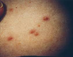
In multisystem disease, bone lesions are still the most common finding, although cutaneous involvement is seen in about one third of patients.
Skin lesions may include dry, scaling skin on the scalp, erythematous rashes in intertriginous regions, or diffuse, papular rashes on the trunk and extremities.
Activated bone marrow cells which are antigen presenting cells that express CD1a and S-100 protein.
The disease is characterized by the presence of large mononuclear cells that normally involved in androgen presenting function and/or CD1a, CD68, S100, and CD207 positive at immunohistochemistry.
It’s pathological cells are found to involve an almost universal activation of the MAPK/ERK pathway, with mutations identified in most kinases upstream of ERK (RAS/RAF/MEK).
Clinically ranges from a self healing to life-threatening illness,characterized by proliferation of myeloid precursors of dendritic cells with ultrastructural and immunophenotypic features of epidermal Langherhans cells.
Langerhans cell histiocytosis involved in a environment made up of T cells, macrophages, and eosinophils causing a local production of cytokines.
LCH is associated accumulation of dendritic cells.
Incidence of 9 cases per million per year.
Affects 1-2 adults and 5-9 children per million.
Usually a pediatric disorder, but adults occasionally affected.
In adults, involvement occurs between the fifth-seventh decades of life both genders equally affected.
Typically limited the involvement of a single bone.
Adult LCH commonly involves bones and maybe a bone limited disease or component of multi system disease, 38% and 66%, respectively.
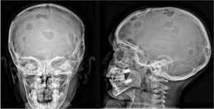
The skull is the most commonly involved, followed by the axial and proximal appendicular skeleton.
Bone involvement usually manifests as pain or as a mass of the involved site.
Aggressive x-ray patterns of bone involvement are more common with moth eaten permeative and cortically based lytic lesions that may progress and are prone to fracture.
PET-CT has superior detection of bone lesions compared to radiographs CT or MRI.
Bone scan his little roll in imaging for LCH.
Considered a hematological disease.
Clinical presentation varies according to organ or tissue involvement.
Can be a local, multifocal or multiply systemic process.
In adults, skin limited forms a rare as most patients with cutaneous involvement display localized disease in internal organs.
Cutaneous manifestations occur in nearly 40% of patients.
Cutaneous disease preferentially localizes to areas of the skin where there is a high production of sebum and typically occurs at flexure sites.
In adults disease related to the skin generally has a better outcome than multi system forms of disease, although patient with cutaneous disease may develop systemic involvement and have a higher risk of secondary hematologic malignancy, mostly Hodgkin’s disease.
Involvement of the genitalia and mucous membranes is common.
More extensive involvement associated with a lower survival rate.
Main form known as eosinophilic granuloma and only affects bones.
Radiographic changes demonstrate osteolytic lesions.
Hand-Schuller-Christian disease (HSC) Is another form of LCH disease and includes diabetes insipidus, exophthalmos, and lytic bone lesions-known as the HSC triad.
Central diabetes insipidus maybe the most important component of HSC as it may be the earliest manifestation, and can precede other manifestations of the disease by years.
Hyperprolactinemia may be present in HSC disease.
Pituitary stalk thickening is an important sign in Hand-Shuller-Christian disease.
Regardless of the organ involved, diagnosis needs to be confirmed by histopathologic and immunohistochemical analyses, and eventually by detecting Birbeck granules through electronic microscopy.
More than 50% of patients have the BRAF V600E mutation.
Localized disease can be treated with low dose radiation therapy.
Patients are stratified high and low risk groups by appropriate therapy.
Local therapy surgical excision, intralesional injection and radiation.
The rarity of disease and variation in presentation makes management controversial.
LCH associated with BRAF V600E mutations in 57% of patients.
Therapeutic options include: surgery, radiation, vinblastine, prednisone, 6-MP, cladribine, methotrexate and cytosine, combination chemotherapy and stem cell transplantation.
BRAF mutation is present in approximately 50% of patients with LCH and BRAF inhibitors are a considered treatment.
Adult patients with multi system disease are candidates for chemotherapy that include Vinblastine regimens.
Adult type diseases associated with long-term morbidity and an overall mortality rate of 3%
In adult patients with skin limited disease less aggressive approaches with oral methotrexate, 6MP, topical steroids, topical nitrogen mustard, or thalidomide have been effective.
The current standard of care for multisystem disease, in patients who have high relapse rate, continues to be a combination of vinblastine and prednisone.
Recommendations for follow up include laboratory testing every six months with blood counts, hormonal and metabolic panel, LDH, beta2 microglobulin and ultrasound of the abdomen and chest x-rays.