1089
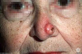
An epithelial neoplasm that is sharply demarcated nodule with the central keratin field crater.
Associated with rapid growth in the proliferative phase and has a variable period of lesion stability.
Has potential for spontaneous involution.
Most commonly seen in patients in their 40s to 60s.
Male to female ratio is 2:1.
Condition is most commonly seen in fair skinned individuals.
Believed to be derived from the infundibulum of the hair follicle.
Gene change expression involving epidermal cells proliferation, cell adhesion and cell survival play a role in its development.
Mutations of the tumor suppressor gene p53 and apoptosis regulatory proteins gene BCL-2/Bak are implicated in its pathogenesis.
Mutations in the transforming growth factor beta receptors and mismatch repair genes have been reported.
Ultraviolet radiation is a predisposing factor.
Predisposing factors include Caucasians with skin photo types I, II, or III, genetic predisposition, trauma, immunodeficiency medications, chemical carcinogens and certain human papilloma virus infections.
Histological findings include epidermal hyperplasia, central keratin field crater, keratinocytes with glassy eosinophilic cytoplasm, sharp demarcation between tumor nests and surrounding stroma, mix inflammatory infiltrate in the dermis.
Lesion does not extend into the dermis below the eccrine glands.
A number of variants have been described: solitary keratoacanthoma, giant keratoacanthoma, subungual keratoacanthoma, mucosal keratoacanthoma, keratoacanthoma centrifugum marginatum, multiple keratoacanthoma syndromes.
Giant keratoacanthoma are greater than 2 cm in diameter and these lesions can be locally invasive and destructive.
Subungual keratoacanthoma develops in the nailbed is usually aggressive and painful
Subungual keratoacanthoma may involve the underlying bone, and be locally aggressive, but does not metastasize.
Mucosal keratoacanthoma develops on surfaces such as the oral cavity, lip, conjunctiva, nasal cavity, and genitalia.
Keratoacanthoma centrifugum marginatum associated with peripheral expansion, raised rolled margin, central healing, and atrophy.
Keratoacanthoma centrifugum marginatum associated with lesions of the dorsum of the hands and feet.
Keratoacanthoma centrifugum marginatum lesions are large up to 20 cm in size, and can be differentiated from giant keratoacanthoma by absence of downward vertical growth and destruction of underlying tissue.
Keratoacanthoma of Grzybowski characterized by sudden onset and erruption of hundreds of thousands of small 1-3 mm follicular, skin colored papules with central umbilication that may contain a central horny, keratotic plug.
Keratoacanthoma of Grzybowski has an onset between the fifth and sixth decades and affects the skin and less commonly the mucous membranes.
Sun exposed skin is a predominantly affected in keratoacanthoma of Grzybowski.
Diagnosis is usually made clinically-based on distinctive history.
Dermoscopy of the lesion reveals keratin crust/scale, central keratin mass, keratin pearls, white structureless central hemorrhage, and areas of keratinization, and atypical blood vessels.
To distinguish squamous cell carcinoma from keratoacanthoma requires a biopsy.
Differential diagnosis includes: squamous cell carcinoma, basal cell carcinoma, Merkel cell carcinoma, actinic keratosis, seborrheic keratoses, verucca vulgaris, pyogenic granuloma, prurigo nodularis, giant molluscum contagiosum, fungal infections and nodular, Kaposi’s sarcoma.
Occasionally such lesions may metastasize.
In approximately 50% of cases spontaneous resolution occurs within 4-9 months of maximal size.
Subungual, mucosal lesions, keratoacanthoma centrifugum marginatum lesions generally do not regress.
Recurrence rate is 3-5%.
Management includes observation, waiting for the lesions to spontaneously resolve.
Many experts believe the lesion is a variant of squamous cell carcinoma and that it should be treated rather than to wait for spontaneous resolution.
Treatment depends on individualization depending on treatment options, the type of keratoacanthoma that is present, its, location, the number and size of lesions and patient preference.
For solitary lesions surgical excision is the treatment of choice.
Other treatment options including electrodissection and curettage, cryosurgery, intralesional methotrexate, topical 5-FU, topical imiquimod or some combination.
For multiple lesions treated options include oral retinoids, oral cyclophosphamide and superficial radiation.
To prevent recurrence avoidance of sun exposure, use of sunscreens and protective clothing should be considered.