It is is composed mainly of either type III, early, or type I late, collagen.
Keloid is a result of an overgrowth of granulation tissue, collagen type 3, at the site of a healed skin injury which is then slowly replaced by collagen type 1.
During wound healing agingEngrailed-1 (EN1) is expressed in fibroblasts of the deep dermis, resulting in a fibrotic response.
The increased tensile forces in a wound reduces the conversion of EN1-lineage- negative fibroblasts to EN1-lineage-positive fibroblasts that result in scarring.
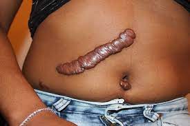
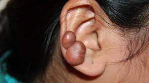
They are rubbery, shiny lesions of fibrous nodules, and can vary from pink to the color of the person’s skin or red to dark brown in color.
A keloid scar is benign.
They are not contagious.
They are sometimes accompanied by severe itchiness, pain, and changes in texture.
In severe cases keloids can affect movement of skin.
In the United States keloid scars are seen 15 times more frequently in people of sub-Saharan African descent than in people of European descent.
Worldwide, men and women of African, Asian, or Hispanic descent can develop keloids.
Some people have a higher tendency to develop a keloid when they scar: family history of keloids and people between the ages of 10 and 30 years.
Keloids form within scar tissue.
Collagen tends to overgrow in areas of wound repair producing a lump that is many times larger than that of the original wound.
Keloids can range in color from pink to red.
Keloids usually occur at the site of an injury, and they can also arise spontaneously.
They can occur at the site of a piercing, a pimple or scratch, severe acne or chickenpox scarring, tattoo, infection at a wound site, repeated trauma to an area, excessive skin tension during wound closure or a foreign body in a wound.
They can sometimes be sensitive to chlorine.
A keloid can grow in someone growing.
Keloids usually develop in any site where skin trauma has occurred: pimples, insect bites, scratching, burns, or other skin injury, as with surgery.
Extensive burns, either thermal or radiological, can lead to unusually large keloids.
Keloids are especially common in firebombing casualties, and were a signature effect of the atomic bombings of Hiroshima and Nagasaki.
Keloids more common in some sites: central chest, the back, shoulders ear lobes, and body piercings.
The most common spots for keloids are earlobes, arms, pelvic region, and over the collar bone.
Types of injuries contributing to scarring: burns, acne scars, chickenpox scars, ear piercing, scratches, surgical incisions, and vaccination sites.
Keloid scarring is common in young people between the ages of 10 and 20.
Individuals with darker complexions are at a higher risk of keloid scarring as a result of skin trauma.
They occur in 15 – 20% of individuals with sub-Saharan African, Asian or Latino ancestry, significantly less in those of a Caucasian background.
Keloids have a genetic component.
Several susceptibility gene loci have been discovered, most notably in Chromosome 15.
People from Sub Saharan Africa, Asia, or Latin America are more likely to develop a keloid.
For Chinese in Asia, the keloid is the most common skin condition.
In the United States, keloids are more common in African Americans and Hispanic Americans than whites.
People of African descent have increased risk of keloid occurrences.
Patients with a family history of keloids are also susceptible: about 1/3 of people who get keloids have a first-degree blood relative who also gets keloids.
Keloids tend to have a genetic component, which means one is more likely to have keloids if one or both of their parents has them.
However, no single gene has yet been identified which is a causing factor in keloid scarring but several susceptibility loci have been discovered, most notably in Chromosome 15.
Family history of keloids is most common in people of African and/or Asian descent.
Development of keloids among twins also lends credibility to genetic susceptibility to develop keloids.
Clinically severe forms of keloids are seen in individuals with a positive family history and black African ethnic origin.
Histologically, they are fibrotic tumors characterized by a collection of atypical fibroblasts.
There are excessive deposits of extracellular matrix components, especially collagen, fibronectin, elastin, and proteoglycans in keloid tissue.
The centers of keloids are relatively acellular with thick, collagen bundles that form nodules in the deep dermal portion of the lesion.
Keloids may be associated with pain, itching, physical disfigurement, and can limit mobility if located over a joint.
Keloids must not improve in appearance over time and can limit mobility if located over a joint.
Keloids affect both sexes equally.
The frequency of occurrence is 15 times higher in highly pigmented people.
Prevention of keloid scars in patients with a known predisposition: by preventing trauma or surgery, ear piercing and elective mole removal, treating skin problems in predisposed individuals with acne, and skin infections.
Prevention of scarring is limited to tension reduction across the wound.
Types of treatments of both preventive and therapeutic nature:
Pressure therapy, silicone gel sheeting, intra-lesional triamcinolone acetonide, cryosurgery, radiation, laser therapy, IFN, 5-FU and surgical excision as well as a extracts and topical agents.
Pressure therapy following surgical excision is effective, especially in keloids of the ear and earlobe.
Radiotherapy, anti-metabolites and corticosteroids would not be recommended to be used in children.
Cryotherapy is associated with least chance of recurrence.
Surgical excision is the most common treatment for a significant amount of keloid lesions.
By itself, surgery has recurrence rate of between 70 and 100%, and can cause a larger lesion on recurrence.
Surgical excision when combined with other therapies dramatically decreases the recurrence rate:
Pressure therapy.
Intralesional injection with a corticosteroid aids in the reduction of fibroblast activity, inflammation and pruritus.
Tea tree oil, salt or other topical oil has no effect on keloid lesions.
Topical oil has no effect on keloid lesions.
Treatment of keloids scar is somewhat age-dependent: radiotherapy, anti-metabolites and corticosteroids would not be recommended to be used in children.
While persons of any age can develop a keloid, children under 10 are less likely to develop keloids, even from ear piercing.
Keloids can occur as a result of severe acne or chickenpox scarring, infection at a wound site, repeated trauma to an area, excessive skin tension during wound closure or a foreign body in a wound.
Keloids may also develop from Pseudofolliculitis barbae.
The tendency to form keloids is suggested to be hereditary.
They can grow over time without even piercing the skin.
Burns, either thermal or radiological, can lead to unusually large keloids, as seen in firebombing casualties, and were a signature effect of the atomic bombings of Hiroshima and Nagasaki.
Reorted incidence of keloids in the general population ranges from a high of 16% among the adults in Zaire to a low of 0.09% in England.
Among sub-Saharan Africans, African Americans and Asians, estimated prevalence rates ranging from 4.5-16%.
Development of keloids among twins also lends credibility to existence of a genetic susceptibility to develop keloids.
Histology: Thick, hyalinized collagen fibers are characteristic, with atypical fibroblasts and excessive deposition of extracellular matrix components, especially collagen, fibronectin, elastin, and proteoglycans.
Keloid tissue contains relatively acellular centers and thick, abundant collagen bundles that form nodules in the deep dermal portion of the lesion.
Keloids affect both sexes equally.
The incidence in young female patients has been reported to be higher than in young males, probably reflecting the greater frequency of earlobe piercing among women.