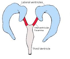
 The lateral ventricles connected to the third ventricle by the interventricular foramina (Foramen of Monro).
In the brain, the interventricular foramina (or foramina of Monro) are channels that connect the paired lateral ventricles with the third ventricle at the midline of the brain.
These channels allow cerebrospinal fluid (CSF) produced in the lateral ventricles to reach the third ventricle and then the rest of the brain’s ventricular system.
The walls of the interventricular foramina also contain choroid plexus, a specialized CSF-producing structure, that is continuous with that of the lateral and third ventricles above and below it.
The choroid plexus, a specialized structure that produces cerebrospinal fluid.
The choroid plexus of the third ventricles continues through the foramina into the lateral ventricles.
The interventricular foramina connect the left and the right lateral ventricles to the third ventricle.
The interventricular foramina are located on the underside near the midline of the lateral ventricles, and join the third ventricle where its roof meets its anterior surface.
In front of the foramen is the fornix and behind is the thalamus.
The foramen is normally crescent-shaped, but rounds out and increases in size depending on the size of the lateral ventricles.
The walls of the interventricular foramina contain choroid plexus, a specialized structure that produces cerebrospinal fluid.
The choroid plexus of the third ventricles continues through the foramina into the lateral ventricles.
The interventricular foramina give rise to disease when they are narrowed or blocked.
Narrowing of the foramen is more common in children.
Narrowing of the interventricular foramen Is linked to: inflammation and scarring from congenital infections, particularly TORCH infections
developmental abnormalities, including of the basilar artery and choroid plexus; and abnormal surrounding tissue growths, such as colloid cysts, subependymal giant-cell tumours, nodules and harmatomas.
The most common symptom of interventricular blockage is headache.
Interventricular blockage symptoms also include’ fainting, dementia, and coma, associated with obstructive hydrocephalus.
Hydrocephalus identified by a CT scan or MRI scan of the brain, can be treated with a neurosurgical operation in which an endoscope is used to widen the foramen or create a new opening through the septum pellucidum between the lateral ventricles.
If an obstructing mass is too large or too difficult to remove endoscopically, an open operation or the insertion of an artificial path between the ventricles and peritoneum may be required.
Such procedures may result in damage to nearby structures, with complications including: amnesia, inability to move half the body, akinetic mutism and disconnection syndromes.