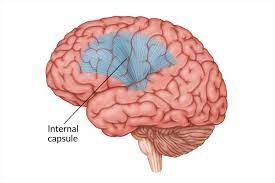
The internal capsule is a bundle of nerve fibers in the brain that connects the cerebral cortex (the outer part of the brain responsible for consciousness) to the brainstem and spinal cord.
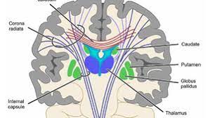
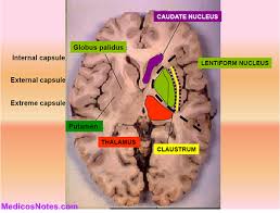
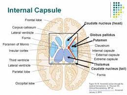
It is located deep within the brain, between the thalamus and the basal ganglia.
The internal capsule (IC) is a white matter structure situated in the inferomedial part of each cerebral hemisphere of the brain.
It carries information past the basal ganglia, separating the caudate nucleus and the thalamus from the putamen and the globus pallidus.
The internal capsule is composed of several distinct regions that carry different types of nerve fibers.
The IC contains both ascending and descending axons, going to and coming from the cerebral cortex.
It also separates the caudate nucleus and the putamen in the dorsal striatum, a brain region involved in motor and reward pathways.
The corticospinal tract constitutes a large part of the internal capsule.
The corticospinal tract carries motor information from the primary motor cortex to the lower motor neurons in the spinal cord.
Above the basal ganglia the corticospinal tract is a part of the corona radiata.
Below the basal ganglia the tract is called cerebral crus, a part of the cerebral peduncle, and below the pons it is referred to as the corticospinal tract.
The internal capsule consists of three parts and is V-shaped when cut horizontally, in a transverse plane.
The bend in the V is called the genu.
The genu is the flexure of the internal capsule, and it is formed by fibers from the corticonuclear tracts.
These geniculate fibers originate in the motor part of the cerebral cortex and after passing downward through the base of the cerebral peduncle with the cerebrospinal fibers, undergo decussation and end in the motor nuclei of the cranial nerves of the opposite side.
The geniculate fibers contains the corticobulbar tract, which carries upper motor neurons from the motor cortex to cranial nerve nuclei that mainly govern motion of striated muscle in the head and face.
The anterior limb of the internal capsule contains fibers that connect the frontal lobe to the thalamus, while the posterior limb contains fibers that connect the occipital lobe to the thalamus and brainstem.
The anterior limb of internal capsule, or frontal part, contains:
fibers running from the thalamus to the frontal lobe
fibers connecting the lentiform and caudate nuclei
fibers connecting the cortex with the corpus striatum
fibers passing from the frontal lobe through the medial fifth of the base of the cerebral peduncle to the nuclei pontis
thalami pontine fibers
The posterior limb of internal capsule (or occipital part) is the portion of the internal capsule posterior to the genu.
The genu of the internal capsule contains fibers that connect the frontal lobe to the basal ganglia.
These nerve fibers within the internal capsule are responsible for transmitting sensory and motor information between the brain and the rest of the body.
The anterior two-thirds of the occipital part of the internal capsule contains fibers of the corticospinal tract, which arise in the motor area of the cerebral cortex and, passing downward through the middle three-fifths of the base of the cerebral peduncle, are continued into the pyramids of the medulla oblongata.
The posterior third of the occipital part contains:
sensory fibers, largely derived from the thalamus, though some may be continued upward from the medial lemniscus
the fibers of optic radiation, from the lower visual centers to the cortex of the occipital lobe;
acoustic fibers, from the lateral lemniscus to the temporal lobe
fibers that pass from the occipital and temporal lobes to the pontine nuclei
The superior parts of both the anterior and posterior limbs and the genu of the internal capsule are supplied by the lenticulostriate arteries, which are branches of the M1 segment of the middle cerebral artery.
The inferior half of the anterior limb is supplied via the recurrent artery of Heubner, which is a branch of the anterior cerebral artery.
The inferior half of the posterior limb is supplied by the anterior choroidal artery, which is a branch of the internal carotid artery.
Anterior limb: lenticulostriate branches of middle cerebral artery.
Genu: lenticulostriate branches of middle cerebral artery.
Posterior limb: lenticulostriate branches of middle cerebral artery and anterior choroidal artery branch of the internal carotid artery.
Some degree of variation in the blood supply exists.
The internal capsule provides passage to ascending and descending fibres running to and from the cerebral cortex.
The anterior limb of the internal capsule contains:
1) Frontopontine fibers project from frontal cortex to the pons;
2) Thalamocortical radiations are the fibers that connect the medial and anterior nuclei of the thalamus to the frontal lobes
The genu contains corticobulbar fibers, which run between the cortex and the brainstem.
The posterior limb of the internal capsule contains corticospinal fibers, sensory fibers from the body and a few corticobulbar fibers.
Other fibers within the internal capsule:
The retrolenticular part contains fibers from the optic system, coming from the lateral geniculate nucleus of the thalamus.
Some fibers from the medial geniculate nucleus carrying auditory information also pass in the retrolenticular internal capsule, but most are in the sublenticular part.
The sublenticular part contains fibers connecting with the temporal lobe.
The lenticulostriate arteries supply a substantial amount of the internal capsule. These small vessels are particularly vulnerable to narrowing in the setting of chronic hypertension and can result in small, punctate infarctions or intraparenchymal haemorrhage due to vessel rupture.
Lesions of the genu of the internal capsule affect fibers of the corticobulbar tract.
The primary motor cortex sends its axons through the posterior limb of the internal capsule.
Lesions in the posterior limb of the internal capsuleresult in a contralateral hemiparesis or hemiplegia.
Damage to the internal capsule can cause a variety of neurological deficits, depending on which fibers are affected: weakness or paralysis of the limbs, sensory loss, and difficulties with speech, language, or cognition.