 The inner ear plays a vital role in the sense of hearing and balance.
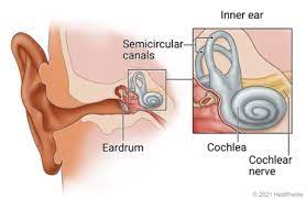
The inner ear is the innermost part of the ear.
It is is mainly responsible for sound detection and balance.
It consists of several structures, including the cochlea, vestibule, and semicircular canals.
It consists of the bony labyrinth, a hollow cavity in the temporal bone of the skull with a system of passages comprising two main functional parts:
The cochlea, dedicated to hearing; converting sound pressure patterns from the outer ear into electrochemical impulses which are passed on to the brain via the auditory nerve.
The vestibular system, dedicated to balance.
The bony labyrinth, or osseous labyrinth, is the network of passages with bony walls lined with periosteum.
The inner ear is innervated by the eighth cranial nerve.
The cochlea is responsible for converting sound vibrations into electrical signals that can be interpreted by the brain.
It contains thousands of tiny hair cells that are sensitive to different frequencies of sound.
These hair cells transmit electrical signals to the auditory nerve, which then sends them to the brain for processing.
The vestibule detects changes in head position and linear acceleration.
The vestibule contains sensory cells and small particles called otoliths, which move in response to gravity or changes in head position.
When the otoliths move, they stimulate the sensory cells and provide information about the body’s orientation.
The semicircular canals are three fluid-filled tubes that are responsible for detecting rotational movements of the head.
Each canal aligns with a different plane of movement, allowing the brain to sense changes in head rotation and help maintain balance.
The inner ear is crucial for the ability to hear and maintain balance.
It works in conjunction with other parts of the ear and the brain to provide us with a comprehensive sensory experience of the world around us.
The membranous labyrinth runs inside of the bony labyrinth, and creates three parallel fluid filled spaces.
The two outer are filled with perilymph and the inner with endolymph.
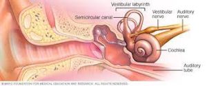
In the middle ear, the energy of pressure waves is translated into mechanical vibrations by the three auditory ossicles.
Pressure waves move the tympanic membrane which in turns moves the malleus, the first bone of the middle ear.
The malleus articulates to incus which connects to the stapes.
The footplate of the stapes connects to the oval window, the beginning of the inner ear.
When the stapes presses on the oval window, it causes the perilymph, the liquid of the inner ear to move.
The middle ear thus serves to convert the energy from sound pressure waves to a force upon the perilymph of the inner ear.
Because the oval window has only approximately 1/18 the area of the tympanic membrane it produces a higher pressure.
The cochlea propagates these mechanical signals as waves in the fluid and membranes and then converts them to nerve impulses which are transmitted to the brain.
The vestibular system is the region of the inner ear where the semicircular canals converge, close to the cochlea.
The vestibular system works with the visual system to keep objects in view when the head is moved.
Joint and muscle receptors are also important in maintaining balance.
The brain receives, interprets, and processes the information from all these systems to create the sensation of balance.
The vestibular system of the inner ear is responsible for the sensations of balance and motion using the same kinds of fluids and detection cells (hair cells) as the cochlea uses, and sends information to the brain about the attitude, rotation, and linear motion of the head.
The type of motion detected by a hair cell depends on its associated mechanical structures, such as the curved tube of a semicircular canal or the calcium carbonate crystals (otolith) of the saccule and utricle.
The spiral canal of the cochlea is a section of the bony labyrinth of the inner ear that is approximately 30 mm long and makes 2¾ turns about the modiolus, the central axis of the cochlea that contains the spiral ganglion.
Inner ear cells include: hair cells, pillar cells, Boettcher’s cells, Claudius’ cells, spiral ganglion neurons, and Deiters’ cells (phalangeal cells).
The hair cells are the primary auditory receptor cells: known as auditory sensory cells, acoustic hair cells, auditory cells or cells of Corti.
The organ of Corti is lined with a single row of inner hair cells and three rows of outer hair cells.
The hair cells have a hair bundle at the apical surface of the cell, consisting of an array of actin-based stereocilia.
Disruption of these bundles results in hearing impairments and balance defects.
Inner and outer pillar cells in the organ of Corti support hair cells.
Both types of pillar cells provide mechanical coupling between the basement membrane and the mechanoreceptors on the hair cells.
Boettcher’s cells are found in the organ of Corti where they are present only in the lower turn of the cochlea, and are supporting cells for the auditory hair cells in the organ of Corti.
Claudius’ cells are found in the organ of Corti located above rows of Boettcher’s cells, are also considered supporting cells for the auditory hair cells in the organ of Corti.
Claudius’ cells contain a variety of aquaporin water channels and appear to be involved in ion transport.
Deiters’ cells.or phalangeal cells, are a type of neuroglial cell found in the organ of Corti and are the supporting cells of the hair cell area within the cochlea.
Hensen’s cells are high columnar cells that are directly adjacent to the third row of Deiters’ cells.
Nuel’s spaces refer to the fluid-filled spaces between the outer pillar cells, the adjacent hair cells and outer hair cells.
The bony labyrinth of the inner ear receives its blood supply from three arteries: 1 – Anterior tympanic branch of the maxillary artery). 2 – Petrosal branch from middle meningeal artery, and 3 – Stylomastoid branch from posterior auricular artery.
The membranous labyrinth is supplied by the labyrinthine artery.
The venous drainage of the inner ear is through the labyrinthine vein.
Neurons within the ear respond to simple tones.
The brain serves to process other increasingly complex sounds.
An average adult is typically able to detect sounds ranging between 20 and 20,000 Hz.
The ability to detect higher pitch sounds decreases in older humans.
The basilar membrane located within the inner-ear resembles that of a traveling wave, the shape of which varies based on the frequency of the pitch: in low-frequency sounds, the tip of the membrane moves the most, while in high-frequency sounds, the base of the membrane moves most.
Infection of the labyrinth can result in a syndrome of ailments called labyrinthitis.
Labyrinthitis symptoms include temporary nausea, disorientation, vertigo, and dizziness.
Labyrinthitis can be caused by viral infections, bacterial infections, or physical blockage of the inner ear.
Autoimmune inner ear disease (AIED) is characterized by idiopathic, rapidly progressive, bilateral sensorineural hearing loss.
The inner ear can detect both static and dynamic equilibrium.
Three semicircular ducts and two chambers, which contain the saccule and utricle, enable the body to detect any deviation from equilibrium.
The macula sacculi detects vertical acceleration while the macula utriculi is responsible for horizontal acceleration.
These microscopic structures possess stereocilia and one kinocilium which are located within the gelatinous otolithic membrane.
The membrane is further weighted with otoliths.
Movement of the stereocilia and kinocilium enable the hair cells of the saccula and utricle to detect motion.
The semicircular ducts are responsible for detecting rotational movement.