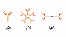
 Immunoglobulin M (IgM) is the largest of several isotypes of antibodies, known as immunoglobulin, that are produced.
IgM is the first antibody to appear in the response to initial exposure to an antigen; called an acute phase antibody.
Plasmablasts in the spleen are the main source of specific IgM production.
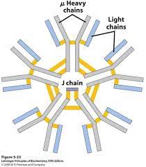
Immunoglobulins are composed of light chains and heavy chains.
The light chain (λ or κ) is a protein of ~220 amino acids, composed of a variable domain, VL (a segment of approximately 110 amino acids), and a constant domain, CL (also approximately 110 amino acids long).
The µ heavy chain of IgM is a protein of ~576 amino acids, includes a variable domain (VH ~110 amino acids), four distinct constant region domains (Cµ1, Cµ2, Cµ3, Cµ4, each ~110 amino acids) and a tailpiece of ~20 amino acids.
The µ heavy chain bears oligosaccharides at five asparagine residues.
The predominant form of IgM is the pentamer.
IgM can bind complement component C1 and activate the classical pathway, which leads to opsonization of antigens and lysis of cells.
IgM binds to mucosal surfaces, such as the gut lumen and into breast milk.
When IgG is administered together with xenogenic erythrocytes, this combination causes almost complete suppression of the erythrocyte-specific antibody response.
This effect is used clinically to prevent Rh-negative mothers from becoming immunized against fetal Rh-positive erythrocytes, and its use has dramatically decreased the incidence of hemolytic disease in newborns.
In contrast to the effect of IgG, antigen-specific IgM can greatly enhance the antibody response, especially in the case of large antigens.
IgM is the first immunoglobulin expressed in the human fetus, which occurs around 20 weeks.
IgM antibodies appear early in the course of an infection and usually reappear, to a lesser extent, after further exposure.
IgM antibodies do not pass across the human placenta, but isotype IgG does.
IgM antibodies in a patient’s serum indicates recent infection, and when present in a neonate’s serum indicates intrauterine infection.
Anti-donor IgM after organ transplantation is not associated with graft rejection.
IgM antibodies are mainly responsible for the clumping or agglutination of red blood cells if the recipient of a blood transfusion receives blood that is not compatible with their blood type.
A mutation of the mu chain within IgM causes autosomal recessive agammaglobulinemia.