 The
The 
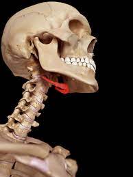
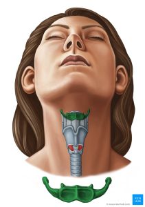
 hyoid bone is a horseshoe-shaped bone located in the anterior midline of the neck, just above the Adam’s apple or laryngeal prominence.
It does not directly articulate with any other bone, but is instead suspended in the neck by muscles and ligaments.
The hyoid bone plays an important role in supporting the muscles of the tongue and the larynx, and it also assists in swallowing and speech production.
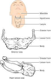
The hyoid bone, at the front of the neck, has a body and two sets of horns
It is situated in the anterior midline of the neck between the chin and the thyroid cartilage.
It is only distantly articulated to other bones by muscles or ligaments.
It is the only bone in the human body that is not connected to any other bones nearby.
The hyoid is anchored by muscles from the anterior, posterior and inferior directions, and aids in tongue movement and swallowing.
The hyoid bone provides attachment to the muscles of the floor of the mouth and the tongue above, the larynx below, and the epiglottis and pharynx behind.
The hyoid bone is an irregular bone and consists of a central part called the body, and two pairs of horns, the greater and lesser horns.
The anterior surface gives insertion to the geniohyoid muscle.
Below the transverse ridge the mylohyoid, sternohyoid, and omohyoid are inserted.
The largest of muscles that attach to the upper surface of the greater horns are the hyoglossus and the middle pharyngeal constrictor, which extend along the whole length of the horns; the digastric muscle and stylohyoid muscle have small insertions in front of these near the junction of the body with the horns.
The lesser horns are two small, conical eminences, connected to the body of the bone by fibrous tissue, and occasionally to the greater horns by distinct diarthrodial joints, which usually persist throughout life, but occasionally become ankylosed.
Blood is supplied to the hyoid bone via the lingual artery, which runs down from the tongue to the greater horns of the bone.
The suprahyoid branch of the lingual artery runs along the upper border of the hyoid bone and supplies blood to the attached muscles.
The hyoid bone allows a wider range of tongue, pharyngeal and laryngeal movements by bracing these structures alongside each other in order to produce variation.
A large number of muscles attach to the hyoid:
Superior
Middle pharyngeal constrictor muscle
Hyoglossus muscle
Genioglossus
Intrinsic muscles of the tongue
Suprahyoid muscles
Digastric muscle
Stylohyoid muscle
Geniohyoid muscle
muscle
Inferior
Thyrohyoid muscle
Omohyoid muscle
Sternothyroid muscle
The hyoid bone is important to a number of physiological functions, including breathing, swallowing and speech.
The hyoid bone plays a key role in keeping the upper airway open during sleep.
The involvement of the hyoid bone is demonstrated to have an inferior position that is strongly associated with the presence and severity of the obstructive sleep apnea.
A surgical procedure to potentially increase and improve the airway is called hyoid suspension.
The hyoid bone is not easily susceptible to fracture.
A fractured hyoid strongly indicates throttling or strangulation.
In children and adolescents the hyoid bone is still flexible because ossification is yet to be completed, a fracture may not occur even after serious trauma.
