Lined with endothelium.
Consists of dense collagenous core near the outflow surface and continuous with valvular supporting structures, a central core of connective tissue and a layer of elastin in the inflow surface.
Collagen in valves responsible for mechanical integrity, the connective tissue absorbs shock of systole, and elastin contracts as the valve cusps are enlarged during diastole.
Interstitial cells in valves produce and repair extracellular matrix.
Leaflets and cusps have minimal vascularization as they are thin enough to receive perfusion from the heart itself.
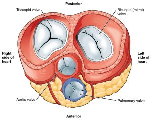
Semilunar valves are the aortic and pulmonary valves.
Competent leaflets (cusps) of the semilunar valves depend on their stretching and molding capabilities to fill the orifice in the closed phase of the cardiac cycle (diastole) when backpressure from the aorta or pulmonary arteries exist.
During the closed phase cusps overlap beneath their free edge.
The semilunar valve leaflets stretch 40-50% larger than their area during systole, the open phase, when they are relaxed.
Function of semilunar depend on the integrity and coordination of the movements of the cusp attachments such that dilation of the aortic root hinders the coaptation of the aortic valve cusps during closure and yields regurgitation.
Aortic root dilation can impair coaptation of the aortic valve cusps during closure and lead to regurgitation of the blood flow.
Pulmonary valve has structure and function similar to the aortic valve.
The atrioventricular valves promote the transit of blood into the ventricles during diastole and prevent regurgitation during systole.
The right heart functioning under lower pressures, ejects the same stroke volume as the high pressure left heart, but in the presence of a closed mitral valve thus relying on the compliance of the pulmonary vasculature and left atrium.
Atrioventricular valves, the mitral and tricuspid, have their free margins tethered to the ventricular wall by many cords, the chordae tendineae, which are attached to papillary muscles contiguous with underlying ventricular walls.
Mitral valve competency dependent on coordination of the annulus, leaflets, cords, papillary muscles and the left ventricle wall to maintain leaflet coaptation.
The left ventricle papillary muscles are positioned beneath the commissures an receive cords from two adjacent leaflets.
Dilation of the left ventricle, rupture of a cord or papillary muscle impair mitral closure causing regurgitation of flow.
Tricuspid valve function is dependent on the same structures as the mitral valve.
Changes that occur in heart valves include damage to the collagen, development of calcifications and fibrotic thickening.
With advanced age many individuals develop small filiform proceses on the closure lines of the aortic and mitral valves, known as Lambl excrescences.
Lambl excrescences probably arise from small thrombi on the valve margins.