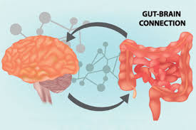
The gut–brain axis refers to the biochemical signaling that takes place between the gastrointestinal tract (GI tract) and the central nervous system (CNS).
The spinal and vagal afferents from the GI tract indirectly project to the thalamus, insula, amygdala, prefrontal cortex, primary somatosensory cortex, secondary somatosensory cortex, and cingulate cortices, including the anterior cingulate cortex.
The brain-gut axis is the mechanism in which the psychosocial factors influence the GI tract and vice versa.
The gut–brain axis is made up of the central nervous system, neuroendocrine and neuroimmune systems, including the hypothalamic–pituitary–adrenal axis (HPA axis), sympathetic and parasympathetic arms of the autonomic nervous system, including the enteric nervous system and the vagus nerve, and the gut microbiota.
The gut–brain axis, is a bidirectional neurohumoral communication system, is important for maintaining homeostasis and is regulated through the central and enteric nervous systems and the neural, endocrine, immune, and metabolic pathways, and especially including the hypothalamic–pituitary–adrenal axis (HPA axis).
The gut-brain axis includes the role of the gut flora as part of the microbiome-gut-brain axis, a linkage of functions including the gut flora.
The gut flora is the complex community of microorganisms that live in the digestive tract.
The gut microbiota has the largest quantity of bacteria and the greatest number of species, compared to other areas of the body.
The gut flora is established at one to two years after birth: the intestinal epithelium and the intestinal mucosal barrier that it secretes have co-developed in a way that is tolerant to, and even supportive of, the gut flora and that also provides a barrier to pathogenic organisms.
Gut microorganisms collecting the energy from the fermentation of undigested carbohydrates and the absorption of short-chain fatty acids (SCFAs), acetate, butyrate, and propionate.
Intestinal bacteria help synthesize vitamin B and vitamin K as well as metabolizing bile acids, sterols, and xenobiotics.
The short chain fatty acids absorbed, and other compounds they produce are like hormones suggest the gut flora itself appears to function like an endocrine organ.
The dysregulation of the gut flora is correlated with a host of inflammatory and autoimmune conditions.
The composition of human gut flora changes over time, when the diet changes, and as overall health changes.
The bioactive compounds, indole and certain other derivatives, from tryptophan are produced by bacteria in the gut.
Indole is produced from tryptophan by bacteria that express tryptophanase.
Clostridium sporogenes metabolizes tryptophan into indole and subsequently 3-indolepropionic acid (IPA).
3-indolepropionic acid is a highly potent neuroprotective antioxidant that scavenges hydroxyl radicals.
IPA binds to intestinal cells, facilitating mucosal homeostasis and barrier function.
Following absorption and distribution to the brain, IPA confers a neuroprotective effect against cerebral ischemia and Alzheimer’s disease.
Lactobacillus species metabolize tryptophan into indole-3-aldehyde (I3A) which acts on intestinal immune cells, in turn increasing interleukin-22 (IL-22) production.
Indole itself triggers the secretion of glucagon-like peptide-1 (GLP-1) in intestinal L cells and acts as a ligand for acetylcholine receptor.
Indole can also be metabolized by the liver into indoxyl sulfate, a compound that is toxic in high concentrations and associated with vascular disease and renal dysfunction.
Activated charcoal, an intestinal sorbent that is taken by mouth, adsorbs indole.
Activated charcoal decreases the concentration of indoxyl sulfate in blood plasma.
The ((enteric nervous system)) consists of a mesh-like system of neurons that governs the function of the gastrointestinal system.
The enteric nervous system can operate autonomously, and normally communicates with the central nervous system (CNS) through the parasympathetic (e.g., via the vagus nerve) and sympathetic (e.g., via the prevertebral ganglia) nervous systems.
The enteric nervous system includes efferent neurons, afferent neurons, and interneurons, all of which make the enteric nervous system capable of carrying reflexes in the absence of CNS input.
The enteric sensory neurons report on mechanical and chemical conditions.
Through intestinal muscles, the motor neurons control peristalsis and churning of intestinal contents.
Additional neurons control the secretion of enzymes.
The enteric nervous system also makes use of more than 30 neurotransmitters, most of which are identical to the ones found in CNS, such as acetylcholine, dopamine, and serotonin.
More than 90% of the body’s serotonin lies in the gut, as well as about 50% of the body’s dopamine.
The beginning of the gut–brain interactions occurs between the sight and smell of food and the release of gastric secretions, known as the cephalic phase, or cephalic response of digestion.
Astrocytes become activated by low glucose and this activation increases gastric emptying to increase digestion.
The gut flora can produce a range of neuroactive molecules, such as acetylcholine, catecholamines, γ-aminobutyric acid, histamine, melatonin, and serotonin, which are essential for regulating peristalsis and sensation in the gut.
Changes in the composition of the gut flora due to diet, drugs, or disease correlates with levels of circulating cytokines, some of which can affect brain function.
The gut flora releases molecules that can directly activate the vagus nerve, which transmits information about the state of the intestines to the brain.
Chronic or acutely stressful situations activate the hypothalamic–pituitary–adrenal axis.
Such stress causes changes in the gut flora and intestinal epithelium, and possibly having systemic effects.
The cholinergic anti-inflammatory pathway, that signals via the vagus nerve, affects the gut epithelium and flora.
Hunger and satiety are integrated in the brain.
The presence or absence of food in the gut and types of food present also affect the composition and activity of gut flora.
Gut microbiota and oral probiotics have been found to influence systemic inflammation, oxidative stress, glycemic control, tissue lipid content, and mood.
People with anxiety and mood disorders tend to have gastrointestinal problems.
Gut flora can produce noradrenalin, and serotonin, that might affect anxiety.
There is a link between the gut microbiome, mood disorders and anxiety, and sleep.
The microbial composition of the gut changes depending on the time of day, so that the gut is exposed to varying metabolites produced by the microbes active during that time.
Microbial changes are associated with differences in the transcription of circadian clock genes involved in circadian rhythm.
Stress and sleep disturbances can lead to greater gut mucosal permeability via activation of the HPA axis, and this causes immune inflammatory responses that contribute to the development of illnesses that cause depression and anxiety.
Around 70% of people with autism also have gastrointestinal problems, and autism is often diagnosed at the time that the gut flora becomes established, indicating that there may be a connection between autism and gut flora.