 Glia, also called glial cells or neuroglia, are non-neuronal cells in the central nervous system of the brain and spinal cord, and the peripheral nervous system that do not produce electrical impulses.
The neuroglia make up more than one half the volume of neural tissue in our body.
Glial cells maintain homeostasis, form myelin in the peripheral nervous system, and provide support and protection for neurons.
In the central nervous system, glial cells include oligodendrocytes, astrocytes, ependymal cells, and microglia, and in the peripheral nervous system they include Schwann cells and satellite cells.
The four types of glial cells have four main functions:
to surround neurons and hold them in place
to supply nutrients and oxygen to neurons
to insulate one neuron from another
to destroy pathogens and remove dead neurons.
Glial cells play a role in neurotransmission and synaptic connections, and in physiological processes such as breathing.
Glial cells have far more cellular diversity and functions than neurons.
Glial cells can respond to and manipulate neurotransmission in many ways, and can affect both the preservation and consolidation of memories.
In the nervous system, non-neuronal cell types, most notably astroglia and microglia, are the primary producers of APOE, while neurons preferentially express the receptors for APOE.
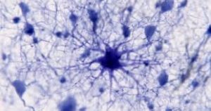
Astrocytes express glial fibrillary acidic protein (GFAP)
Astrocytes are the most abundant type of macroglial cell in the CNS.
Astrocytes have numerous projections that link neurons to their blood supply while forming the blood–brain barrier.
Astrocytes regulate the external chemical environment of neurons by removing excess potassium ions, and recycling neurotransmitters released during synaptic transmission.
Astrocytes may regulate vasoconstriction and vasodilation by producing substances such as arachidonic acid, whose metabolites are vasoactive.
Astrocytes signal each other using ATP.
The gap junctions between astrocytes allow the messenger molecule IP3 to diffuse from one astrocyte to another.
IP3 activates calcium channels on cellular organelles, releasing calcium into the cytoplasm.
This calcium may stimulate the production of more IP3 and cause release of ATP through channels in the membrane made of pannexins.
The net effect is a calcium wave that propagates from cell to cell.
Extracellular release of ATP, and consequent activation of purinergic receptors on other astrocytes, may also mediate calcium waves in some cases.
In general, there are two types of astrocytes, protoplasmic and fibrous, similar in function but distinct in morphology and distribution.
Protoplasmic astrocytes have short, thick, highly branched processes and are typically found in gray matter.
Fibrous astrocytes have long, thin, less branched processes and are more commonly found in white matter.
Astrocyte activity is linked to blood flow in the brain, and that this is what is actually being measured in fMRI.
Astrocytes also have been involved in neuronal circuits playing an inhibitory role after sensing changes in extracellular calcium.
Oligodendrocytes are cells that coat axons in the central nervous system (CNS) with their cell membrane, forming a specialized membrane differentiation called myelin, producing the myelin sheath.
The myelin sheath provides insulation to the axon that allows electrical signals to propagate more efficiently.
Ependymal cells, also named ependymocytes, line the spinal cord and the ventricular system of the brain.
These cells are involved in the creation and secretion of cerebrospinal fluid (CSF) and beat their cilia to help circulate the CSF and make up the blood-CSF barrier.
They are also thought to act as neural stem cells, and function both as neuronal progenitors and as a scaffold upon which newborn neurons migrate.
In the mature brain, the cerebellum and retina retain characteristic radial glial cells.
Schwann cells provide myelination to axons in the peripheral nervous system (PNS).
They also have phagocytotic activity and clear cellular debris that allows for regrowth of PNS neurons.
Satellite glial cells are small cells that surround neurons in sensory, sympathetic, and parasympathetic ganglia, helping to regulate the external chemical environment.
Satellite glial cells like astrocytes, they are interconnected by gap junctions and respond to ATP by elevating the intracellular concentration of calcium ions.
Satellite glial cells are highly sensitive to injury and inflammation and contribute to pathological states, such as chronic pain.
Enteric glial cells are found in the intrinsic ganglia of the digestive system.
Glia cells are thought to have many roles in the enteric system, some related to homeostasis and muscular digestive processes.
Microglia are specialized macrophages capable of phagocytosis that protect neurons of the central nervous system.
Microglia are found in all regions of the brain and spinal cord.
They are small relative to macroglial cells, with changing shapes and oblong nuclei.
They are mobile within the brain.
Microglia multiply when the brain is damaged.
Microglia processes constantly sample their environmental neurons, macroglia and blood vessels.
Microglia direct the immune response to brain damage and play an important role in the inflammation that accompanies such damage.
Many diseases and disorders are associated with deficient microglia, such as Alzheimer’s disease, Parkinson’s disease, and ALS.
Pituicytes from the posterior pituitary are glial cells with characteristics in common to astrocytes.
In general, neuroglial cells are smaller than neurons.
There are approximately 85 billion glia cells in the human brain, about the same number as neurons.
Glial cells make up about half the total volume of the brain and spinal cord.
The glia to neuron-ratio varies from one part of the brain to another.
The glia to neuron-ratio in the cerebral cortex is 3.72 (60.84 billion glia (72%); 16.34 billion neurons).
The ratio in the cerebellum is only 0.23 (16.04 billion glia; 69.03 billion neurons).
The ratio in the cerebral cortex gray matter is 1.48, with 3.76 for the gray and white matter combined.
The ratio of the basal ganglia, diencephalon and brainstem combined is 11.35.
The total number of glia cells in the human brain is distributed into the different types with oligodendrocytes being the most frequent (45–75%), followed by astrocytes (19–40%) and microglia (about 10% or less).
Most glia are derived from ectodermal tissue of the developing embryo, mainly the neural tube and crest.
Microglia, however, are derived from hemopoietic stem cells.
In the adult, microglia are largely a self-renewing population and are distinct from macrophages and monocytes, which infiltrate an injured and diseased CNS.
In the central nervous system, glia develop from the ventricular zone of the neural tube,and include the oligodendrocytes, ependymal cells, and astrocytes.
In the peripheral nervous system, glia derive from the neural crest.
These PNS glia include Schwann cells in nerves and satellite glial cells in ganglia.
Glia retain the ability to undergo cell divisions in adulthood, whereas most neurons cannot.
There is an inability of the mature nervous system to replace neurons after an injury, such as a stroke or trauma, but there is a substantial proliferation of glia, or gliosis, near or at the site of damage.
There is no evidence that mature Glia, such as astrocytes or oligodendrocytes, retain mitotic capacity, but resident oligodendrocyte precursor cells seem to keep this ability once the nervous system matures.
Some glial cells function primarily as the physical support for neurons.
Some glial cells provide nutrients to neurons and regulate the extracellular fluid of the brain, especially surrounding neurons and their synapses.
During early embryogenesis, glial cells direct the migration of neurons and produce molecules that modify the growth of axons and dendrites.
Some glial cells display regional diversity and their functions may vary between the CNS regions.
Glia are vital in the development of the nervous system and in synaptic plasticity and synaptogenesis, and have a role in the regulation of repair of neurons after injury.
In the central nervous system (CNS), glia suppress repair, as astrocytes enlarge and proliferate to form a scar and produce inhibitory molecules that inhibit regrowth of a damaged or severed axon.
In the peripheral nervous system glial cells known as Schwann cells promote repair.
After axonal injury, Schwann cells regress to an earlier developmental state to encourage regrowth of the axon.
Oligodendrocytes have bulbous cell bodies with up to fifteen arm-like processes.
Each process reaches out to an axon and spirals around it, creating a myelin sheath.
The myelin sheath insulates the nerve fiber from the extracellular fluid and speeds up signal conduction along the nerve fiber.
In the peripheral nervous system, Schwann cells are responsible for myelin production.
These cells envelop nerve fibers of the PNS by winding repeatedly around them, creating a myelin sheath, which not only aids in conductivity but also assists in the regeneration of damaged fibers.
Astrocyte functions, include clearance of neurotransmitters from within the synaptic cleft, which aids in distinguishing between separate action potentials and prevents toxic build-up of certain neurotransmitters such as glutamate, which would otherwise lead to excitotoxicity.
Furthermore, astrocytes release gliotransmitters such as glutamate, ATP, and D-serine in response to stimulation.
Gial cells in the PNS frequently assist in regeneration of lost neural functioning, but loss of neurons in the CNS does not result in a similar reaction from neuroglia.
In the CNS, regrowth will only happen if the trauma was mild, and not severe.
Glial cells affect the potential repair of neurons in Alzheimer’s disease, scarring and inflammation from glial cells have been further implicated in the degeneration of neurons caused by amyotrophic lateral sclerosis.
When damage occurs to the CNS, glial cells cause apoptosis among the surrounding cellular bodies.
Increased microglial activity results in inflammation, and finally, and a heavy release of growth inhibiting molecules.
Albert Einstein’s brain was discovered to contain significantly more glia than normal brains in the left angular gyrus, an area thought to be responsible for mathematical processing and language.
The number of glial cells in the brain is correlated with the intelligence of a species, and the active role of glia, in particular astroglia, in cognitive processes like learning and memory.