A fetus is the unborn offspring that develops from an embryo.
In human prenatal development, fetal development begins from the first week after fertilization and continues until birth.
A fetus receives an estimated 3 to 4 g per kilogram of body weight of amino acids per day supporting fetal growth and brain development.
There is no clear defining feature distinguishing an embryo from a fetus.
A fetus is characterized by the presence of all the major body organs, though not yet be fully developed and functional.
In humans, the fetal stage starts nine weeks after fertilization.
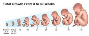
At the start of the fetal stage, the fetus is typically about 30 millimetres (1+1⁄4 in) in length from crown-rump, and weighs about 8 grams.
The head makes up nearly half of the size of the fetus.
Breathing-like movements of the fetus are necessary to stimulate lung development, rather than for obtaining oxygen.
The heart, hands, feet, brain and other organs are present, but are only at the beginning of development and have minimal operation.
At this point in development, uncontrolled movements and twitches occur as muscles, the brain, and pathways begin to develop.
Weeks 17 to 25 or 3.6 to 6.6 months
A woman pregnant for the first time typically feels fetal movements at about 21 weeks, whereas a woman who has given birth before will typically feel movements by 20 weeks.
By the end of the fifth month, the fetus is about 20 cm (8 in) long.
Weeks 26 to 38 or 6.6 to 8.6 months.
The amount of body fat rapidly increases.
Lungs are not fully mature.
At 30 weeks gestation the sensory cortex and thalamus neural connections develop and minimal consciousness, dreaming, and the ability to feel pain emerges.
Bones are fully developed at this point, but are still soft and pliable.
Iron, calcium, and phosphorus become more abundant in bones.
Fingernails reach the end of the fingertips at this time.
Lanugo, which are fine hair, begins to disappear, except on the upper arms and shoulders.
Small breast buds are present on both fetal sexes.
Head hair becomes coarse and thicker.
Imminent birth occurs around the 38th week after fertilization.
The fetus is considered full-term between weeks 36 and 40.
A fetus may be 48 to 53 cm (19 to 21 in) in length, when born.
Control of movement is limited at birth.
Variations in the growth of the human fetus occur.
When fetal size is less than expected, the condition is known as intrauterine growth restriction (IUGR) also called fetal growth restriction (FGR).
Factors affecting fetal growth can be due to maternal, placental, or fetal matters.
Maternal factors include: maternal weight, body mass index, nutritional state, emotional stress, toxin exposures to tobacco, alcohol, heroin, and other drugs which can also harm the fetus, and uterine blood flow.
Placental factors include: its size, microstructure, blood flow, transporters and binding proteins, nutrient utilization and nutrient production.
Fetal factors include the fetus genome, nutrient production, and hormone output.
Female fetuses tend to weigh less than males, at full term.
Fetal growth is characterized as follows: small for gestational age (SGA), appropriate for gestational age (AGA), and large for gestational age (LGA).
Low birth weight increases risk for perinatal mortality, asphyxia, hypothermia, polycythemia, hypocalcemia, immune dysfunction, neurologic abnormalities, and other long-term health problems.
SGA may be associated with growth delay, or an absolute stunting of growth.
Fetal viability refers to a point in fetal development at which the fetus may survive outside the womb.
The lower limit of fetal viability is approximately 5+3⁄4 months gestational age, but there is no sharp limit of development, age, or weight at which a fetus automatically becomes viable.
Survival rates are 20–35% for babies born at 23 weeks of gestation (5+3⁄4 months); 50–70% at 24–25 weeks (6 – 6+1⁄4 months); and >90% at 26–27 weeks (6+1⁄2 – 6+3⁄4 months) and over.
For a baby weighing less than 500 g (1 lb 2 oz) survival is rare.
The main causes of mortality in highly premature babies are that the respiratory system and the central nervous system are not completely differentiated.
Preterm birth is the most common cause of infant mortality, causing almost 30 percent of neonatal deaths.
Preterm birth rate is 5% to 18% of all deliveries.
Preterm birth is more common than postmature birth, which occurs in 3% to 12% of pregnancies.
The fetus obtains oxygen and nutrients from the mother through the placenta and the umbilical cord.
The heart and blood vessels of the circulatory system, form relatively early during embryonic development, and
are biological necessary, since mammalian tissues can not grow more than a few cell layers thick without an active blood supply.
The prenatal circulation of blood is different from postnatal circulation, mainly because the lungs are not in use.
Blood from the placenta is carried to the fetus by the umbilical vein.
About half of the umbilical vein blood flow enters the fetal ductus venosus and is carried to the inferior vena cava, while the other half enters the liver proper from the inferior border of the liver.
The umbilical vein that supplies the right lobe of the liver joins with the portal vein.
The blood carried to the inferior vena cava moves to the right atrium of the heart.
In the fetus, the opening between the right and left atrium, the foramen ovale, causes most of the blood flow from the right into the left atrium, thus bypassing pulmonary circulation.
The majority of blood flow in the fetal circulation flows into the left ventricle from where it is pumped through the aorta into the body.
Blood that moves from the aorta through the internal iliac arteries to the umbilical arteries, re-enters the placenta, where carbon dioxide and other waste products from the fetus are taken up and enter the mother’s circulation.
Some of the blood from the right atrium does not enter the left atrium, but enters the right ventricle and is pumped into the pulmonary artery.
The fetus, has a special connection between the pulmonary artery and the aorta, the ductus arteriosus, which directs most of this blood away from the lungs.
The lungs are not used by the fetus for respiration, as the fetus is suspended in amniotic fluid.
The first breath after birth causes system changes.
Resistance i the lungs is reduced dramatically, allowing more blood to move into the pulmonary arteries from the right atrium and ventricle of the heart and less to flow through the foramen ovale into the left atrium.
The blood from the lungs travels through the pulmonary veins to the left atrium, producing an increase in pressure that pushes the septum primum against the septum secundum, closing the foramen ovale and completing the separation of the newborn’s circulatory system into the standard left and right sides.
The foramen ovale is known subsequently as the fossa ovalis.
The ductus arteriosus normally closes within one or two days of birth, leaving the ligamentum arteriosum.
The umbilical vein and ductus venosus usually closes within two to five days after birth, leaving the liver’s ligamentum teres and ligamentum venosus, respectively.
The placenta functions as a maternal-fetal barrier against the transmission of microbes.
Maternal IgG antibodies cross the placenta.
This gives the fetus passive immunity against those diseases for which the mother has antibodies.
This transfer of antibodies to the fetus begins as early as the fifth month of gestational age.
A developing fetus is highly susceptible to anomalies in its growth and metabolism.
Supplementation of the person’s diet with folic acid reduces the risk of spina bifida and other neural tube defects.
It is suggested skipping breakfast could lead to extended periods of lower than normal nutrients in the maternal blood, leading to a higher risk of prematurity, or birth defects.
Alcohol consumption may cause fetal alcohol syndrome, a condition leading to intellectual disability in some infants.
Smoking during pregnancy may lead to miscarriages and low birth weight.
X-ray radiation is known to have possible adverse effects on the development of the fetus, and the risks need to be weighed against the benefits.
Infants with certain congenital heart defects can survive only as long as the ductus remains open, and closure of the ductus can be delayed by the administration of prostaglandins to permit sufficient time for the surgical correction of the anomalies.
In patent ductus arteriosus, where the ductus does not properly close, drugs that inhibit prostaglandin synthesis can be used to encourage its closure, to avoid surgery.
Fetal perception of pain is unlikely before the third trimester.
However, the establishment of thalamocortical connections at about 6+1⁄2 months is an essential event with regard to fetal perception of pain.
Abortion of a human pregnancy is legal and/or tolerated in most countries, although with gestational time limits that normally prohibit late-term abortions.