
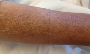
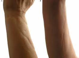
 Eosinophilic fasciitis is a rare connective tissue disorder within the scleroderma spectrum, characterized by inflammation and fibrosis of the deep fascia.
It typically presents with acute or subacute painful swelling (edema) and progressive thickening (induration) of the limbs, often with a “peau d’orange” (orange peel) appearance.
This condition is frequently associated with peripheral eosinophilia, hypergammaglobulinemia, and elevated inflammatory markers.
Diagnosis is best confirmed by a full-thickness biopsy that includes the skin, fascia, and muscle, revealing fascial thickening with inflammatory infiltrates.
Fibrosis and tethering of connective tissue around superficial veins, cause the venous groove sign, a linear depression, resulting from overlying epidermis being pulled inward along a vein.
MRI can also be useful in supporting and guiding the evaluation.
This inflammatory disease primarily affects the fascia, but can also involve other connective tissues, as well as surrounding muscles, blood vessels, and nerves.
Unlike other forms of fasciitis, eosinophilic fasciitis is typically self-limited and confined to the arms and legs, although treatment with corticosteroids may be necessary.
Some cases are also associated with aplastic anemia.
Eosinophilic fasciitis is associated with hematologic malignancies-Hodgkin disease, CLL, multiple myeloma, graft versus host disease, hemodialysis initiation, radiation therapy, and sustained intense physical exertion.
Certain medications, including immune checkpoint inhibitors, statins, and proton pump inhibitors have been reported to be associated with eosinophilia fasciitis
The presentation of eosinophilic fasciitis is similar to scleroderma or systemic sclerosis, but unlike scleroderma, eosinophilic fasciitis affects the deeper fascial layers.
Eosinophilic fasciitis is a very rare disease, with only about 100 cases reported.
The typical age of onset is between 20 to 60 years, though cases in children have been observed. It is slightly more common in men than in women, with a ratio of 1.5:1.
Most cases are idiopathic, but some have been associated with:
Strenuous exercise Initiation of hemodialysis Infection with Borrelia burgdorferi Certain medications, such as statins, phenytoin, ramipril, and subcutaneous heparin
Other autoimmune diseases, like systemic sclerosis, systemic lupus erythematosus, and Sjögren syndrome
Hematologic disorders like aplastic anemia, multiple myeloma, chronic lymphocytic leukemia, and other leukemias and lymphomas
The main symptoms include pain and swelling in the distal extremities.
Presentation usually involves symmetrical swelling and thickening of the skin on the forearms and lower legs, accompanied by pain and erythema.
The hands and feet are typically spared.
Differential diagnosis: systemic sclerosis, scleraomyxedema, and scleredema, and eosinophilia myalgia syndrome.
Treatment:
First line treatment is oral steroids.
Immunotherapy suppressant therapy such as methotrexate and micophenolate may be added for extensive disease or to help taper or discontinue steroids.
Tocilizumab, baricitinib, infliximab, rituximab and intravenous immunoglobulin may also be effective.
Physical therapy to prevent contract contractions as recommended.