 Endometriosis is a chronic, estrogen-dependent gynecologic disease defined as the presence of endometrial-like epithelial and/or stromal tissue outside the uterine cavity and myometrium.
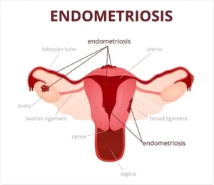
These ectopic implants are most frequently found in the pelvic peritoneal cavity, but can occur anywhere, including ovaries, rectovaginal septum, bowel, diaphragm, pleura, and rarely more distant sites like brain, lung, umbilicus.
Epidemiology It affects ≈ 6–11% of reproductive-age women (~190 million worldwide, ~9–11 million in the US).
Prevalence is 2–11% in asymptomatic women, 20–50% in infertile women, and 50–80% in women with chronic pelvic pain.
Among adolescents with chronic pelvic pain or dysmenorrhea unresponsive to usual therapy, prevalence reaches 49–75%.
Typical age at diagnosis: early 30s, despite symptom onset usually in adolescence/early 20s with diagnostic delay averaging 5–12 years.
Pathogenesis The exact cause is unknown; multiple mechanisms likely coexist: Retrograde menstruation (Sampson’s theory) – most widely accepted for pelvic disease. Coelomic metaplasia. Lymphatic/vascular metastasis explaining extrapelvic sites.
Stem-cell implantation, immune dysfunction, genetic/epigenetic factors.
Endometriotic lesions produce their own estradiol locally, elicit chronic inflammation with cytokine/chemokine/prostaglandin release, recruit macrophages and other immune cells, and promote angiogenesis and neurogenesis.
These processes create a self-sustaining pro-inflammatory, pro-angiogenic, estrogen-rich microenvironment.
Clinical Presentation
Pelvic pain (70–90%): dysmenorrhea, chronic non-cyclic pelvic pain, deep dyspareunia, dyschezia, dysuria. Pain often worsens over time and with menses. Infertility (≈30–50% of patients). Fatigue (moderate–severe in ~50%, independent of anemia or mood/sleep disorders). Cyclic symptoms at extrapelvic sites (catamenial pneumothorax, hemoptysis, umbilical bleeding).
Up to 20–25% of women are asymptomatic.
Pain is multifactorial: nociceptive (inflammation), neuropathic (perineural invasion, nerve sensitization by IGF-1 from macrophages), and nociplastic (central sensitization with altered brain connectivity and glutamatergic neurotransmission).
Physical Examination Often normal. May reveal uterine retroversion/fixation, adnexal mass (endometrioma), uterosacral nodularity or tenderness, or reduced uterine mobility in deep disease.
Diagnosis Gold standard: laparoscopic visualization ± histologic confirmation.
Imaging: Transvaginal ultrasound – first-line; excellent (>90% sensitivity/specificity) for ovarian endometriomas, reasonable for deep nodules. MRI – useful for deep infiltrating endometriosis and when ultrasound is equivocal.
Superficial peritoneal lesions and adhesions are poorly seen on any imaging.
No reliable non-invasive biomarker (CA-125 is non-specific).
Empiric medical therapy without surgical confirmation is now widely accepted given long diagnostic delays.
Classification / Staging American Society for Reproductive Medicine (ASRM) stages I–IV based on location, depth, and adhesions.
Staging correlates poorly with pain severity or fertility outcomes (except deep disease somewhat correlates with dyspareunia).
Subtypes of endometriosis: 1 Superficial peritoneal endometriosis 2 Ovarian endometriomas (“chocolate cysts”) 3 Deep infiltrating endometriosis (>5 mm depth; often rectovaginal, bladder, bowel) 4 Extrapelvic / thoracic / abdominal wall, etc.
Natural History in observational surgical studies, lesions progressed in ~29%, regressed in ~42%, and remained stable in ~29%.
Symptoms generally improve after natural or surgical menopause unless hormone replacement is used.
Treatment Goals
Endometriosis is a chronic condition without medical or surgical cure. Pain relief Preserve/improve fertility when desired Prevent or delay recurrence There is currently no cure.
Medical Management (first-line for pain, not seeking pregnancy)
NSAIDs ± acetaminophen (limited evidence).
Combined oral contraceptives (continuous > cyclic) or progestins (norethindrone acetate, dienogest, medroxyprogesterone, levonorgestrel-IUS) – similar efficacy, well tolerated.
All hormonal treatments target sex steroid dependent pathophysiology of endometriosis by suppressing ovarian activity and creating a low estrogen environment, which can lead to regression of endometrial lesions.
GnRH agonists (leuprolide, goserelin, etc.) or oral GnRH antagonists (elagolix 150–200 mg daily; relugolix + estradiol + norethindrone combination) – second-line; require add-back therapy (low-dose estrogen–progestin) to prevent bone loss.
Aromatase inhibitors (letrozole, anastrozole) ± progestin – third-line for refractory cases (off-label; monitor bone density).
Danazol and older anti-progestins are rarely used due to side-effect profile.
Surgical Management Diagnostic and therapeutic laparoscopy with excision/ablation of lesions + lysis of adhesions, preferred over coagulation alone.
Excision of deep nodules, endometriomas, bowel/bladder resection when indicated.
Hysterectomy ± bilateral salpingo-oophorectomy is definitive for women who have completed childbearing and have severe, refractory symptoms (ovaries should be preserved when possible in premenopausal women).
Recurrence after surgery: ~20–40% within 5 years; pain relief not guaranteed in central sensitization cases.
Fertility Considerations
Surgery may improve spontaneous pregnancy rates in minimal–mild disease; benefit in moderate–severe disease is less clear and ovarian reserve can be harmed.
IVF is often required; endometriosis lowers success rates.
Recurrence and Chronicity
50% of women have symptom recurrence within 5 years regardless of medical or surgical approach. 11–19% are completely refractory to hormonal therapy.
Complications and Associations Slightly increased risk of ovarian cancer (clear-cell and endometrioid subtypes). Higher prevalence of autoimmune diseases, non-Hodgkin lymphoma, melanoma. Significant economic burden (direct + indirect costs ~$70–100 billion annually in the US).
Pain does not correlate well with visible disease burden.
Early empiric hormonal therapy is appropriate while awaiting definitive diagnosis.
Multidisciplinary, individualized, long-term management is required because this is a chronic, relapsing condition with no current cure.