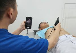
Emergency ultrasound employs point-of-care ultrasound (POCUS) at the point of care to make immediate patient-care decisions.
It is performed by the health care professional caring for the injured or ill persons.
POCUS is often to evaluate an emergency medical condition, in settings such as an emergency department, critical care unit, ambulance, or combat zone.
It is used to quickly diagnose a limited set of injuries or pathologic conditions.
Specifically it is useful where conventional diagnostic methods would either take too long or would introduce greater risk to the patient, as with radiation.
It is now used in various clinical settings at the bedside: In the emergency setting, it is used to guide resuscitation and monitor critically ill persons, provide procedural guidance for improved safety and confirm clinical diagnosis.
Point-of-care ultrasound examinations are performed and interpreted by the same clinician and are typically narrower in scope.
Point of care ultrasound is sometimes the only option in the evaluation of injured persons who are too ill for transport to other imaging modalities.
It is used to guide and triage care in resource-limited situations, in rural or medically under-served areas.
It is used to assess hypotensive persons for occult bleeding.
Traditionally used by emergency physicians and surgeons treating trauma persons, by paramedics in combat zones, and for non-traumatic problems such as ruptured ectopic pregnancy.
Ultrasound can also evaluate the lungs for hemothorax and pneumothorax, to determine the cause of shock, to evaluate the heart and inferior vena cava and can help the clinician at the bedside choose important treatments and monitor the response to the interventions.
An ultrasound showing hyperdynamic left heart with a flat, collapsible IVC indicates low blood volume, it may identify an abdominal aortic aneurysm that is leaking or ruptured, and a cardiac cause for low blood pressure, can assess acute shortness of breath by assessment of the lung, heart, and IVC can evaluate for potentially life-threatening diseases, including pneumothorax, significant pleural effusions, congestive heart failure, pulmonary edema, pericardial effusion, and some large pulmonary emboli.
Ultrasound is now frequently used more in code situations, to see if the heart is moving, beating in organized fashion or if it has a pericardial effusion or fluid around it.
Pericardiocentesis, utilizes ultrasound guidance of a needle to decrease the risk of hitting lungs, heart or other vital organs.
Ultrasound can be utilized to assess a person’s intravascular volume status and response to intravenous fluid therapy by measuring the size and respiratory change in the diameter of the IVC.
It can assess central venous collapsibility as a more standardized measure of intravascular volume status.
It can be used to assess peripheral veins in estimating intravascular volume status in the absence of IVC visualization.
Ultrasound of the lungs may demonstrate resolution of pulmonary edema from congestive heart failure.
Using ultrasound to guide needles during procedures for central and venous access, arterial cannulation, thoracentesis, paracentesis, pericardiocentesis, arthrocentesis, regional anesthesia, incision and drainage of abscesses, localization and removal of foreign bodies, lumbar puncture, and biopsies.
Point-of-care ultrasound can be increasingly used to speed patient care and to avoid ionizing radiation.
Focused cardiac ultrasound can be helpful in the evaluation of persons with potentially life-threatening disease such as a pericardial effusion, a severe pulmonary embolus, screening those with suspected aortic dissection.
Emergency ultrasound of the gallbladder can help speed diagnosis and care of gallbladder disease.
Flank pain can indicate obstructing kidney stones or abdominal aortic aneurysm.
The kidneys can be evaluated by ultrasound for signs of obstruction with called hydronephrosis.
It can identify or evaluating the fetus, or tubal or ectopic pregnancy in a person who is pregnant.
A bedside ultrasound can determine the presence or absence of blood clots and their location in the proximal lower extremity to behind the knee.
An ultrasound of the eye can be used for the detection of orbital pathology: detect retinal detachments, vitreous hemorrhage, dislocation of the lens, as well as evaluating optic nerve sheath diameters as a potential indicator of other diseases in the central nervous system.
The use of point-of-care ultrasound can be used to diagnose appendicitis, testicular torsion, and abscesses.
Evaluation of testicular Pain –testicular torsion, orchitis, Emergency ultrasound
Emergency ultrasound employs point-of-care ultrasound (POCUS) at the point of care to make immediate patient-care decisions.
It is performed by the health care professional caring for the injured or ill persons.
POCUS is often to evaluate an emergency medical condition, in settings such as an emergency department, critical care unit, ambulance, or combat zone.
It is used to quickly diagnose a limited set of injuries or pathologic conditions.
Specifically it is useful where conventional diagnostic methods would either take too long or would introduce greater risk to the patient, as with radiation.
It is now used in various clinical settings at the bedside: In the emergency setting, it is used to guide resuscitation and monitor critically ill persons, provide procedural guidance for improved safety and confirm clinical diagnosis.
Point-of-care ultrasound examinations are performed and interpreted by the same clinician and are typically narrower in scope.
Point of care ultrasound is sometimes the only option in the evaluation of injured persons who are too ill for transport to other imaging modalities.
It is used to guide and triage care in resource-limited situations, in rural or medically under-served areas.
It is used to assess hypotensive persons for occult bleeding.
Traditionally used by emergency physicians and surgeons treating trauma persons, by paramedics in combat zones, and for non-traumatic problems such as ruptured ectopic pregnancy.
Ultrasound can also evaluate the lungs for hemothorax and pneumothorax, to determine the cause of shock, to evaluate the heart and inferior vena cava and can help the clinician at the bedside choose important treatments and monitor the response to the interventions.
An ultrasound showing hyperdynamic left heart with a flat, collapsible IVC indicates low blood volume, it may identify an abdominal aortic aneurysm that is leaking or ruptured, and a cardiac cause for low blood pressure, can assess acute shortness of breath by assessment of the lung, heart, and IVC can evaluate for potentially life-threatening diseases, including pneumothorax, significant pleural effusions, congestive heart failure, pulmonary edema, pericardial effusion, and some large pulmonary emboli.
Ultrasound is now frequently used more in code situations, to see if the heart is moving, beating in organized fashion or if it has a pericardial effusion or fluid around it.
Pericardiocentesis, utilizes ultrasound guidance of a needle to decrease the risk of hitting lungs, heart or other vital organs.
Ultrasound can be utilized to assess a person’s intravascular volume status and response to intravenous fluid therapy by measuring the size and respiratory change in the diameter of the IVC.
It can assess central venous collapsibility as a more standardized measure of intravascular volume status.
It can be used to assess peripheral veins in estimating intravascular volume status in the absence of IVC visualization.
Ultrasound of the lungs may demonstrate resolution of pulmonary edema from congestive heart failure.
Using ultrasound to guide needles during procedures for central and venous access, arterial cannulation, thoracentesis, paracentesis, pericardiocentesis, arthrocentesis, regional anesthesia, incision and drainage of abscesses, localization and removal of foreign bodies, lumbar puncture, and biopsies.
Point-of-care ultrasound can be increasingly used to speed patient care and to avoid ionizing radiation.
Focused cardiac ultrasound can be helpful in the evaluation of persons with potentially life-threatening disease such as a pericardial effusion, a severe pulmonary embolus, screening those with suspected aortic dissection.
Emergency ultrasound of the gallbladder can help speed diagnosis and care of gallbladder disease.
Flank pain can indicate obstructing kidney stones or abdominal aortic aneurysm.
The kidneys can be evaluated by ultrasound for signs of obstruction with called hydronephrosis.
It can identify or evaluating the fetus, or tubal or ectopic pregnancy in a person who is pregnant.
A bedside ultrasound can determine the presence or absence of blood clots and their location in the proximal lower extremity to behind the knee.
An ultrasound of the eye can be used for the detection of orbital pathology: detect retinal detachments, vitreous hemorrhage, dislocation of the lens, as well as evaluating optic nerve sheath diameters as a potential indicator of other diseases in the central nervous system.
The use of point-of-care ultrasound can be used to diagnose appendicitis, testicular torsion, and abscesses.
Evaluation of testicular Pain –testicular torsion, orchitis, epididymitis, hernia.
Evaluation of pelvic pain-ovarian torsion, ovarian cyst, and ovarian cyst rupture
Evaluation of volume status and estimation of fluid responsiveness.’
Exam for Shock or Hypotension
Bones – Evaluation for fractures
Evaluation of pelvic pain-ovarian torsion, ovarian cyst, and ovarian cyst rupture
Evaluation of volume status and estimation of fluid responsiveness.’
Exam for Shock or Hypotension
Bones – Evaluation for fractures