 Electromyography (EMG) is a diagnostic test that records and analyzes the electrical activity of muscles to evaluate neuromuscular disorders.
Electromyography (EMG) measures the electrical activity produced by skeletal muscles and evaluates how well the nerves controlling those muscles are functioning.
It helps detect neuromuscular abnormalities, such as issues with nerve-to-muscle communication, muscle dysfunction, or nerve damage.
It is typically performed alongside nerve conduction studies (NCS) to distinguish between muscle disorders (myopathies), nerve disorders (neuropathies), neuromuscular junction disorders, and motor neuron diseases.
EMG study involves inserting a needle electrode into muscle tissue to record bioelectric signals generated by muscle fibers during rest and contraction.
EMG evaluates spontaneous activity, motor unit action potential (MUAP) morphology, and recruitment patterns.
When combined with NCS, which measure the speed and strength of electrical signals traveling through nerves, the complete electrodiagnostic evaluation provides comprehensive assessment of the neuromuscular system.
EMG testing is indicated for patients presenting with muscle weakness, tingling or numbness in extremities or face, muscle cramps, spasms, twitching, or paralysis.
EMG is typically ordered to investigate to help diagnose or rule out conditions such as: • Carpal tunnel syndrome • Peripheral neuropathies (e.g., diabetic neuropathy) • Pinched nerves (radiculopathy, e.g., from herniated discs) • Muscle disorders (myopathies, muscular dystrophy) • Motor neuron diseases (e.g., ALS) • Myasthenia gravis or other neuromuscular junction disorders
The EMG test helps localize pathology, helps in differential diagnoses, and guide appropriate biopsy sites when needed.
Neuropathies typically show delayed or reduced motor unit activation, spontaneous discharges, and impaired nerve-muscle communication.
Myopathies display low-amplitude signals and rapid motor unit recruitment due to intrinsic muscle weakness.
EMG can help track denervation and reinnervation processes, revealing fibrillation potentials or polyphasic motor unit potentials during nerve recovery.
EMG remains cost-effective for guiding diagnostic workup before ordering expensive genetic panels.
Procedure EMG usually involves two main parts:
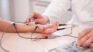
Nerve Conduction Study which is often done first:
Small surface electrodes are placed on the skin.
Mild electrical pulses stimulate nerves, and responses are recorded.
Needle EMG: A thin needle electrode is inserted into specific muscles.
It records electrical activity at rest, during slight contraction, and during stronger effort.
The EMG test usually takes 30–90 minutes, depending on how many muscles/nerves are examined.
Surface electrodes cause minimal sensation..
Generally very safe with rare complications-minor bleeding, infection, or soreness.