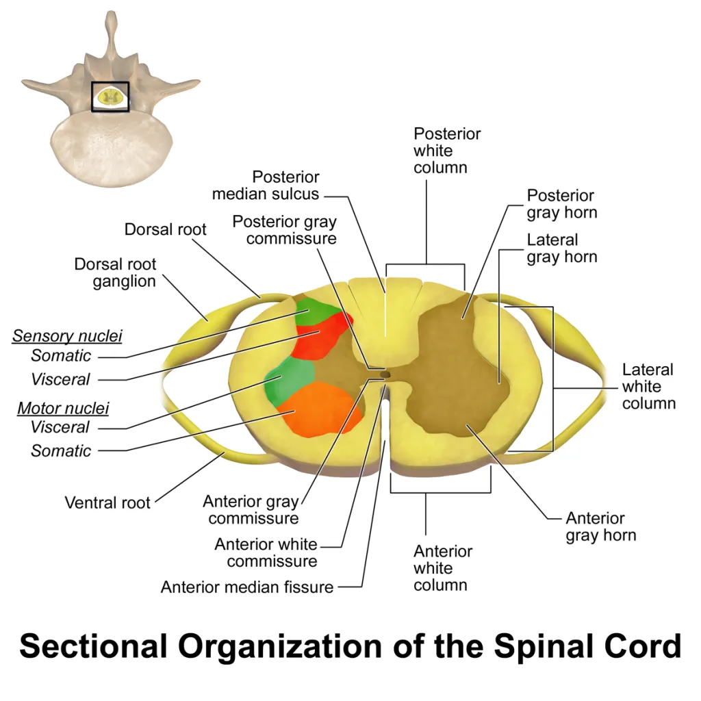
Each dorsal root of the spinal cord contains at the level of the intervertebral foramen an elongated thickening called spinal ganglion or dorsal root ganglion (DRG).
It is a thickening, sheathed by the continuation of the surrounding membranes of the cord into the epineurium and perineurium of the peripheral nerves, and is caused by the accumulation of cell bodies of primary sensory neurons.
A dorsal root ganglion also known as a posterior root ganglion is a cluster of neurons in a dorsal root of a spinal nerve.
Its cell bodies of sensory neurons known as first-order neurons are located in the dorsal root ganglia.
The axons of
Dorsal root ganglion neurons are known as afferent axons.
In the peripheral nervous system, afferent axons relay sensory information into the central nervous system.
The neurons comprising the dorsal root ganglion have a cell body (soma) with two branches that act as a single axon, often referred to as a distal process and a proximal process.
Unlike the majority of neurons found in the central nervous system, an action potential in posterior root ganglion neuron may initiate in the distal process in the periphery, bypass the cell body, and continue to propagate along the proximal process until reaching the synaptic terminal in the posterior horn of spinal cord.
The distal section of the axon may either be a bare nerve ending or encapsulated by a structure that helps relay specific information to nerve.
The nerve ending of the distal process is encapsulated in Meissner’s corpuscles, which render the distal processes of mechanosensory neurons sensitive to stroking only, and Pacinian corpuscles, which make neurons more sensitive to vibration.
The dorsal root ganglia lies in the intervertebral foramina.
The anterior and posterior spinal nerve roots join just lateral to the location of the dorsal root ganglion.
The dorsal root ganglia develop from neural crest cells, and can be regarded as gray matter of the spinal cord that became translocated to the periphery.
The nerve endings of dorsal root ganglion neurons have sensory receptors that are activated by mechanical, thermal, chemical, and noxious stimuli.
In these sensory neurons ion channels are thought to be responsible for somatosensory transduction.
Compression of the dorsal root ganglion by a mechanical stimulus lowers the voltage threshold needed to evoke a response and causes action potentials to be fired.
Two distinct types of mechanosensitive ion channels exist in the posterior root ganglion neurons: high-threshold (HT) or low threshold (LT).
These are cationic channels whose activity appears to be regulated by the cytoskeleton and cytoskeleton associated proteins.
High-threshold channels have a role in nociception, and are found predominantly in smaller sensory neurons in the dorsal root ganglion cells and are activated by higher pressures, two attributes that are characteristic of nociceptors.
GABAA receptors can presynaptically control nociception and pain transmission.
The single axonal process divides in a T-like fashion into a peripheral branch, which is connected to somatic and visceral receptors, and a central branch, which enters the cord.
The dorsal spinal root comprises the trajectory of this axonal bundle between its entry into the cord and the intervertebral foramen.
Distal to the DRG, it fuses with the ventral spinal root to form the spinal nerve.
The more caudal the cord level, the longer the corresponding proximal dorsal root.
The root sheath covers this part of the dorsal root, which traverses the subarachnoid space to the cord, and is considered a continuation of both pia and arachnoid mater but possibly also of the deep perineurium.
The proximal part of each dorsal root of one ganglion is composed of a number of smaller rootlets that enter the dorsolateral cord in a row.
The central projections of a single DRG may also ascend or descend to terminate in neighboring cord segments, thus causing overlap of the spinal segments and of the dermatomal zones.
A large amount of dorsal root fiber collaterals ascend uninterruptedly in the cord through the ipsilateral dorsal column (dorsal funiculus) to terminate in the dorsal column nuclei.
In this respect, the dorsal columns are in fact a central continuation of the peripheral nervous system.