 Hip dysplasia is an abnormality of the hip joint where the socket portion does not fully cover the ball portion, resulting in an increased risk for joint dislocation.
Developmental dysplasia of the hip is one of the most common musculoskeletal conditions in infants and is present in in 4 to 23 infants per 1000 live births.
DDH is a spectrum of that abnormalities ranging from instability of the hip joint to complete dislocation.
Risk factors include female, sex, first-degree family member with DDH, breech presentation during pregnancy, foot deformities, certain swaddling practices, and breech presentation whether an infant is delivered vaginally or by cesarean section.
Native Americans are more likely to have congenital hip dislocation than any of the other races.
The risk for Native Americans is about 25–50 in 1000.
The overall frequency of developmental dysplasia of the hip is approximately 1 case per 1000 individuals.
Some believe that the incidence of hip instability in newborns can be as high as 1 case for every 60 newborns, with the rate dropping to 1:240 at one week.
The condition is eight times more frequent in females than in males.
If one identical twin is affected, there is a 40% risk the other will also be affected.
Hip dysplasia may occur at birth or develop in early life.
Hip dysplasia does not typically produce symptoms in babies less than a year old.
Occasionally one leg may be shorter than the other,and the left hip is more often affected than the right.
Complications of hip dysplasia without treatment include: arthritis, limping, and low back pain.
Females are affected more often than males.
Screening all babies for the condition by physical examination is recommended.
Diagnostic methods:
Physical exam, ultrasound
Treatment
Bracing, casting, surgery.
Prognosis is good if detected and treated early.
Many cases with mild instability resolve without specific treatment.
In more significant cases, if detected early, bracing may be all that is required.
In cases of hip dysplasia that are detected later, surgery and casting may be needed.
About 7.5% of hip replacements are done to treat problems related to hip dysplasia.
Hip instability of meaningful importance occurs in one to two percent of babies born at term.
Females are affected more often than males.
Hip dysplasia can range from barely detectable to severely malformed or dislocated.
The congenital form, teratologic or non-reducible dislocation occurs as part of more complex medical conditions.
The condition can be bilateral or unilateral:
In unilateral dysplasia only one joint shows deformity, the opposite side may show resulting effects.
In the majority of unilateral cases, the left hip has the dysplasia.
If the hip joint is fully dislocated a false acetabulum often forms opposite the dislocated femoral head position.
In acetabular dysplasia, the acetabulum socket is too shallow or deformed.
Two forms of femoral dysplasia are coxa vara, in which the femur head grows at too narrow an angle to the shaft, and coxa valga, in which the angle is too wide.
Hip dysplasia is considered to be a multifactorial condition.
Hip dysplasia cause is unknown; however, some factors of congenital hip dislocation are through heredity and racial background.
Higher rates in some ethnic groups,such as some Native Americans, is due to the practice of swaddling of infants, which is known to be a potential risk factor for developing dysplasia.
Hip dysplasia has a low risk in African Americans.
The hormone relaxin has been implicated in hip dysplasia.
Female sex, alone without other known risk factors, accounts for 75%.
A genetic factor is indicated as the trait runs in families and there is an increased occurrence in some ethnic populations (e.g., Native Americans).
A locus has been described on chromosome 13. and chromosome 4.
Further risk factors include being a firstborn.
The breech position is probably the most important single risk factor, whether an infant is delivered vaginally or by cesarean section.
The acquired condition it has been linked to swaddling infants, use of overly restrictive baby seats, carriers and other methods of transporting babies, or use of a cradle board which locks the hip joint in an adducted position of pulling the knees together tends to pull the heads of the femur bone out of the sockets or acetabulae.
A narrow uterus also facilitates hip joint dislocation during fetal development and birth.
All newborns should be screened for congenital hip dysplasia.
The screening examination techniques detect in hip dysplasia in asymmetry of legs and asymmetrical gluteal folds , and restricted hip abduction.
Two maneuvers commonly employed for diagnosis in neonatal exams are the Ortolani maneuver and the Barlow maneuver.
If a clicking/clucking sound can be heard, it indicates that the baby may have a dislocated hip.
Follow-up exams and developmental monitoring are important.
Physical examination of newborns followed by appropriate use of hip ultrasound is common.
Hip dysplasia can develop in older age.
Adolescents and adults with hip dysplasia may present with a waddling gait, Trendelenburg’s sign, decreased hip abduction, and hip pain
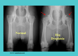
X-rays are used to confirm a diagnosis of hip dysplasia.
CT scans and MRI scans are occasionally used too.
The term DDH encompasses occult dysplasia (e.g. an underdeveloped joint) without dislocation and a dislocation developing after the newborn period.
Types of DDH include subluxation, dysplasia, and dislocation.
The main types are the result of either laxity of the supporting capsule or an abnormal acetabulum.
Hip dysplasia can be diagnosed by ultrasound and projectional X-ray
Ultrasound imaging is generally preferred at up to 4 months due to limited ossification of the femoral head, and is the most accurate method for imaging of the hip during the first few months after birth.
Ultrasound screening should not be performed before 3 to 4 weeks of age because of the normal physiologic laxity.
Despite the widespread use of ultrasound, pelvis X-ray is still frequently used to diagnose or monitor hip dysplasia or for assessing other congenital conditions or bone tumors.
Hip dysplasia presents a conundrum between the arthritis, movement/mobility problems and pain associated with the developmental malformation, and the arthritis, movement/mobility problems and pain that are, as often as not in moderate to severe cases, inflicted by the treatment itself.
The worst consequence of non treatment is developing early arthritis, sometimes even during teenage years.
Treatments aim to delay the onset of arthritis.
No treatment is fully successful in avoiding arthritis, and, all available treatments bear the risk of inflicting equivalent damage.
If diagnosed before an infant is three months of age, a removable hip brace is an effective treatment in 90% of cases.
A delayed diagnosis of after four months of age frequently requires surgery to restore the position of the hip mobilization of the hip and thigh for up to six months in a spica cast.
Studies have as yet been unable to find a method of predicting outcomes in either the surgical/orthopedic treatment of the condition in infants and young children, or the surgical treatment of these early treatments’ negative outcomes later in life,such as arthritis, avascular necrosis, trochanteric bursitis, and bone spurs.
Early hip dysplasia can often be treated using a harness or a pillow/splint in the first year of life with usually normal results.
Complications of femoral nerve palsy and avascular necrosis of the femoral head have been reported with the use of the harness.
Complications occur because the sheet of the iliopsoas muscle pushes the circumflex artery against the neck of the femur and decreases blood flow to the femoral head
Other devices employed include the spica cast.
In older children the adductor and iliopsoas muscles may have to be treated surgically because they adapt to the dislocated joint position by contractures.
Braces and splints are often used following either of these methods.