 A branch of pathology that studies and diagnoses diseases on the cellular level.
Cytopathology is generally used on samples of free cells or tissue fragments, in contrast to histopathology, which studies whole tissues.
Cytopathology is commonly used to investigate diseases involving a wide range of body sites, often to aid in the diagnosis of cancer but also in the diagnosis of some infectious diseases and other inflammatory conditions.
A common application of cytopathology is the Pap smear, a screening tool used to detect precancerous cervical lesions that may lead to cervical cancer.
Cytopathologic tests are sometimes called smear tests because the samples may be smeared across a glass microscope slide for subsequent staining and microscopic examination.
Cytology samples may be prepared in other ways, including cytocentrifugation.
There are two methods of collecting cells for cytopathologic analysis: exfoliative cytology, and intervention cytology.
With exfoliative cytopathology specimen cells are collected after they have been either spontaneously shed by the body (pleural fluid cells) or manually scraped/brushed off of a surface in the body (cervical smear).
Fine-needle aspiration, or fine-needle aspiration cytology (FNAC), involves use of a needle attached to a syringe to collect cells from lesions in various body organs by microcoring.
FNAC can be performed under palpation guidance on a mass in superficial regions like the neck, thyroid or breast; or may be assisted by ultrasound or CAT scan for sampling of deep-seated lesions within the body that cannot be localized via palpation.
Fine needles are 23 to 27 gauge.
FNAC has become synonymous to interventional cytology.
In sediment cytology the sample is collected from the fixative that was used for processing the biopsy specimen and is centrifuged.
The sediment is used for smearing cells that are shed by the biopsy specimen during processing.
Imprint cytology refers to a preparation procedure wherein the tissue of interest touches a glass slide, leaving behind its imprint in the form of cells on the slide.
The imprint can subsequently be stained and studied.
After sampling, two main techniques for processing are used:
Processing of specimens may result in visual artifacts:
Smearing of cells across a glass plate may cause a variety of smearing artifacts.
To better visualize cells and their components, specimens are inked: such Papanicolaou stain, or Romanowsky stain derivatives which include Giemsa, Jenner, Wright, Field, May–Grünwald and Leishman stains.
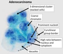
In cancerous cells, altered DNA activity can be seen as a physical change in the nucleus.
As more DNA is unfolded and being expressed, the nucleus is darker and less uniform, larger than in normal cells, and often show a bright-red nucleolus.
Cytopathology can detect if cancerous or precancerous pathology is present in the cellular material, along with other pathologies:
microbial infections: parasitic, viral, and/or bacterial
reactive changes
immune reactions
cell aging
amyloidosis
autoimmune diseases
Cytopathology can detect abnormal functions of cell growth, metabolism, and divisions that lead to diseases.
Cytopathology is used used with physical examination and medical imaging to determine the presence of disease.
Cytology can be used to diagnose a condition and spare a patient from surgery to obtain a larger specimen.
A fine-needle aspiration can be done anywhere it is safe to put a needle, including liver, lung, kidney, and superficial masses.
Cytological specimens must be properly prepared so that the cells are not damaged.
Further information about the specimen may be gained by immunohistochemical stains and molecular testing.
Cytopathologic techniques are used in the examination of virtually all body organs and tissues:
Gynecologic cytology
Urinary tract cytology
Effusion cytology
Breast cytology
Vaginal cytology
Thyroid cytolologu
Lymph node cytology
Respiratory cytology
Gastrointestinal cytology
Soft tissue, bone and skin cytology
Kidney and adrenal cytology
Liver and pancreas cytology
Central nervous system cytology
Eye cytology
Salivary gland cytology