 The cochlea is the sense organ that translates sound into nerve impulses to the brain.
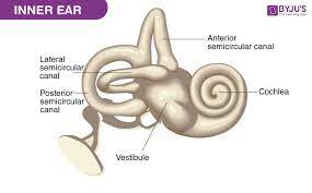
A fluid-filled, snail shaped cavern in the bony part of the skull near each ear.
It is the auditory portion of the inner ear.
It is a spiral-shaped cavity in the bony labyrinth, making 2.5 turns around its axis, the modiolus.
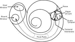
A core component of the cochlea is the Organ of Corti, the sensory organ of hearing, which is distributed along the partition separating fluid chambers in the coiled tapered tube of the cochlea.
Tiny bones in the middle ear transmit sound from the ear drum across the middle ear and vibrate against the cochlea.
Vibrations in the fluid cause tiny hair cells inside the cochlea to vibrate and generate nerve impulses that travel to the brain.
The spiral canal of the cochlea is a section of the bony labyrinth of the inner ear that is approximately 30 mm long and makes 2¾ turns about the modiolus, the central axis of the cochlea that contains the spiral ganglion.
Inner ear cells include: hair cells, pillar cells, Boettcher’s cells, Claudius’ cells, spiral ganglion neurons, and Deiters’ cells (phalangeal cells).
The hair cells are the primary auditory receptor cells: known as auditory sensory cells, acoustic hair cells, auditory cells or cells of Corti.
The organ of Corti is lined with a single row of inner hair cells and three rows of outer hair cells.
The hair cells have a hair bundle at the apical surface of the cell, consisting of an array of actin-based stereocilia.
Disruption of these bundles results in hearing impairments and balance defects.