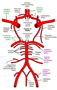
The circle of Willis is a circulatory anastomosis that supplies blood to the brain and surrounding structures.
Blood flows up to the brain through the vertebral arteries and through the internal carotid arteries.
The circle of Willis is a part of the cerebral circulation and is composed of the following arteries:
Anterior cerebral artery
Anterior communicating artery
Internal carotid artery
Posterior cerebral artery
Posterior communicating artery
The middle cerebral arteries, supplying the brain, are not considered part of the circle of Willis.
The left and right internal carotid arteries arise from the left and right common carotid arteries.
The posterior communicating artery is given off as a branch of the internal carotid artery just before it divides into its terminal branches – the anterior and middle cerebral arteries.
The anterior cerebral artery forms the anterolateral portion of the circle of Willis, while the middle cerebral artery does not contribute to the circle.
The right and left posterior cerebral arteries arise from the basilar artery, which is formed by the left and right vertebral arteries.
The vertebral arteries arise from the subclavian arteries.
The anterior communicating artery connects the two anterior cerebral arteries and could be said to arise from either the left or right side.
All arteries involved give off cortical and central branches.
The central branches supply the interior of the circle of Willis, more specifically, the Interpeduncular fossa.
Considerable anatomic variation exists in the circle of Willis.
The classic anatomy of the circle is only seen in 34.5% of cases.
A common variation the proximal part of the posterior cerebral artery is narrow and its ipsilateral posterior communicating artery is large, so the internal carotid artery supplies the posterior cerebrum; known as a fetal posterior communicating cerebral artery.
Another variation the anterior communicating artery is a large vessel, such that a single internal carotid supplies both anterior cerebral arteries; this is known as an azygos anterior cerebral artery.
The circle of Willis creates redundancy for collateral circulation in the cerebral circulation.
If part of the circle becomes blocked or narrowed or one of the arteries supplying the circle is blocked or narrowed, blood flow from the other blood vessels can often preserve the cerebral perfusion well enough to avoid the symptoms of ischemia.
Its other functions: dampening of pulse pressure waves within the brain and involvement in forebrain sensing of water loss.
The adaptive flow that the circle of Willis introduces can also lead to reduced cerebral perfusion, as with the subclavian steal syndrome.
In the subclavian steal syndrome, blood is “stolen” from the vertebral artery on the affected side to preserve blood flow to the upper limb.
The subclavian steal syndrome results from a proximal stenosis of the subclavian artery, one of arteries originating off of the aortic arch, and has potential to affect flow in the circle of Willis.