Chronic venous insufficiency affects 2-5% of Americans.
Chronic venous insufficiency risk factors include older age, female sex, obesity, pregnancy, history of deep vein thrombosis, and prolonged standing.
Additional risk factors associated with the development of varicose veins include low bioimpedance and greater height.
855 single nucleotide, polymorphisms and 30 independent genetic variants are associated with varicose veins, establishing a genetic contribution to their development.
Chronic venous insufficiency of the legs are among the most frequent vascular conditions, affecting millions of people worldwide.
Venous insufficiency increases the risk of venous thrombotic events and is associated with a substantial limitations on daily functioning and quality of life.
Chronic venous disease encompasses the entirety of venous disorder is whereas chronic venous insufficiency denotes the more advanced forms of venous disorders, such as edema, skin, manifestations of hyperpigmentation, and healed or active venous ulcers are present.
Chronic venous insufficiency arises from complex relationships with genetic predisposition, environmental factors such as job requirements of standing/ carrying heavy loads, and age related loss of structural integrity of the venous system in the legs.
Veins are capacitance blood vessels that accommodates approximately 2/3 of the total blood volume in the body.
The venous system serves as a reservoir for the circulatory system, enabling rapid adjustments in volume with varied pressures.
When in the supine position, central venous pressure ranges from 8 to 12 mmHg and could be as high as 90 mmHg in the legs while standing upright.
The venous system comprises both superficial and deep veins.
The deep veins are responsible for greater than 90% of venous return.
Foot and calf muscle contractions can initiate cephalad flow of blood and opens the one way valves within the veins, preventing retrograde flow after closing.
Dysfunctional valves cause venous reflux characterized by backflow of blood, which results in venous hypertension and deleterious affects overtime.
Venous hypertension causes, endothelial dysfunction, increases, vascular, permeability, and induces vein wall inflammatory response.
These conditions result in disruption of the reduction-oxidation balance equilibrium between the generation and elimination of reactive oxygen and nitrogen species leading to tissue degradation and clinical manifestations of varicose veins, edema, skin changes, and venous ulcers.
Blood Venous disorder stages include:
Stage 0: No signs that can be seen or felt.
Stage 1: Visible blood vessels, including spider veins.
Stage 2: Varicose veins at least 3 millimeters wide.
Stage 3: Edema (swelling) but no skin changes.
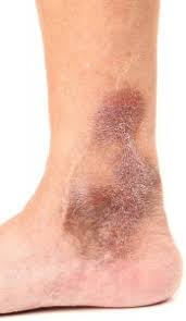
Stage 4: Changes to your skin’s color and/or texture.
Stage 5: Healed ulcer.
Stage 6: Acute (active) ulcer.
Can be caused by congenital absence or damage to the venous valves in superficial and communicating systems.
Can be caused by thrombi formation in veins.
Varicose veins are rarely associated with the development of chronic venous insufficiency.
Venous hypertension is prevented by venous bicuspid valves that prevent backflow and venous blood pooling, and by calf muscle contractions which decreases venous pressures by approximately 70% in the lower extremities.
During rest calf muscles rest and venous pressures return to normal in 30 seconds.
In disease veins ambulation decreases venous pressures by only 20% and when ambulation stops the venous pressure returns to normal over a period of minutes.
Venous hypertension causes chronic venous insufficiency by transcending venous pressure from the venules to the capillaries, impeding blood flow.
When venous pressure impedes blood flow capillary stasis causes leucocyte trapping with release of proteolytic enzymes and oxygen free radicals damaging the capillary basement membranes.
Associated with family history of deep vein thrombophlebitis, sedentary lifestyle, more frequent in obese women, more common
Among those who stand for long periods of time and increased incidence in men who smoke.
Signs include edema, hyperpigmentation of the skin, venous eczema, atrophic changes, and induration of subcutaneous fat by fibrosis.
Seven classes C0-C6 with symptoms (S) or asymptomatic (A).
Symptoms include aching, heaviness, irritation, feeling of swelling.
Chronic venous insufficiency refers to disease of severity classes C4 to C6.
Ankle edema present in 7% of men and 16% of women.
Active or healed venous leg ulcers occur in approximately 1% of general population.
Prevalence, particularly of leg ulcers increases with age.
More prevalent among women.
Accounts for 92 per 100,000 hospital admissions.
Peak incidence in women aged 40-49 years and in men 70-79 years.
Associated with chronic life endangering infections of the lower extremities, pain after ambulating, lipodermatosclerosis of the skin of the lower extremities and skin ulcerations.
Approximately 6 million patients in the U.S. have skin changes associated with chronic venous insufficiency.
Venous stasis ulcers present in approximately 500,000 Americans.
Lifestyle changes and compression therapy are the initial therapy.
Lifestyle changes:
Achieve and maintain a healthy weight to reduce strain on the legs.
Exercise regularly, focusing on activities that improve leg pumping, such as walking and running.
Elevating the legs and short walk breaks if you have to sit for long periods of time
Lifting the legs above the level of your heart can help reduce pressure in your leg veins.
It is suggested to do this for 30 minutes or longer at least three times per day.
Weight management
Compression therapy
Compression therapy helps ease swelling and discomfort in the legs.
There are many types of compression bandages and stockings.
Some stockings are graduated, meaning they’re tighter down at the ankles and less tight further up the leg.
Intermittent pneumatic compression (IPC) devices may be helpful.
Medications used to treat CVI include:
Antibiotics to clear skin infections or ulcers
Anticoagulants, to treat blood clots and prevent future blood clots from forming.
Medicated wrap known as an Unna boot combines multilayer compression with a zinc oxide gel-based wound cover that forms a semi-rigid bandage.
Nonsurgical treatment
Sclerotherapy
Endovenous thermal ablation
Surgical treatment
Surgical treatments for CVI include:
Ligation and stripping
Microincision/ambulatory phlebectomy
Subfascial Endoscopic Perforator Surgery (SEPS):
Vein bypass:
Compression therapy involves the use of special garments that put external pressure on the leg to push valves closer together, increasing their efficiency.
Compression socks and stockings provide support and apply graduated compression.
Sclerotherapy involves injecting chemicals into affected veins to seal them closed and prevent blood from flowing through them.
Over time, the treated veins turn to scar tissue and fade away as the body redirects blood flow to healthier veins.
Radiofrequency ablation involves inserting a thin catheter into the dysfunctional vein.
The end of the catheter is a heating element that heats up and seals off the vein from the inside.
Microphlebectomy for veins that are too small for ablation or too big for sclerotherapy can be treated with this outpatient procedure.
VenaSeal use a catheter to apply a small amount of medical adhesive inside dysfunctional veins; The adhesive closes off these veins, and the body reroutes blood flow to healthy veins over the next few weeks.