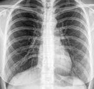
 Also called a chest X-ray (CXR), chest film or chest radiograph.
It is a projection radiograph of the chest used to diagnose conditions affecting the chest, its contents, and nearby structures.
The most common film taken in medicine.
It employs ionizing radiation in the form of X-rays to generate images of the chest.
The mean radiation dose to an adult from a chest radiograph is around 0.02 mSv (2 mrem) for a front view (PA or posterior-anterior) and 0.08 mSv (8 mrem) for a side view (LL or latero-lateral).
Together these views corresponds to a background radiation equivalent time of about 10 days.
Conditions commonly identified by chest X-RAYS:
Pneumonia
Pneumothorax
Interstitial lung disease
Heart failure
Bone fracture
Hiatal hernia
Chest radiographs are used to diagnose: conditions involving the chest wall, including its bones, and also structures contained within the thoracic cavity including the lungs, heart, and great vessels.
Pneumonia and congestive heart failure are very commonly diagnosed by chest radiograph.
Chest x-rays are typically the first test in evaluating chest pain and can exclude pneumothorax, pneumonia, pleural effusion, and pulmonary edema, but lacks specificity and sensitivity for many other processes.
Used to screen for job-related lung disease.
A chest X-ray may identify problems in:
Airways
Breast shadows
Bones
Cardiac silhouette
Costophrenic angles
Diaphragm
Lung Edges
Extrathoracic tissues
Lung fields
The most common views are posteroanterior, anteroposterior, and lateral.
In an posteroanterior (PA) view, the x-ray source is positioned so that the x-ray beam enters through the posterior aspect of the chest, and exits out of the anterior (front) aspect where the beam is detected.
A radiation source is positioned behind the patient at a standard distance, most often 6 feet, and the x-ray beam is fired toward the patient.
In anteroposterior (AP) views, the positions of the x-ray source and detector are reversed: the x-ray beam enters through the anterior aspect and exits through the posterior aspect of the chest.
AP chest x-rays are more difficult to interpret PA x-rays and are reserved for situations where it is difficult for the patient to get an ordinary chest x-ray, such as when the patient is bedridden.
Mobile X-ray equipment is used to obtain a lying down chest x-ray.
Lateral views of the chest are obtained as patient stands with both arms raised and the left side of the chest pressed against a flat surface.
Chest radiography includes a PA and Lateral with the patient standing or sitting up.
Special projections include an anterior posterior, lateral decubitus , axial Lordotic views.
Lateral decubitus may be used for visualization of air-fluid levels if an upright image cannot be obtained.
Axial lordotic views projects the clavicles above the lung fields, allowing better visualization of the apices.
Lordotic views are used to visualize the apex of the lung, to pick up abnormalities such as a Pancoast tumor, or pneumothorax.
Decubitus films are taken while the patient is lying down, typically their side, and is used to differentiate pleural effusions from consolidation and loculated effusions from free fluid in the pleural space.
Decubitus films demonstrate effusions, as the fluid layers out.
Oblique views are useful for the visualization of the ribs and sternum.
Review of CXR it should appear that in the average person, the diaphragm should be intersected by the 5th to 7th anterior ribs at the mid-clavicular line, and 9 to 10 posterior ribs should be viewable on a normal PA inspiratory film.
The presence of an increase in the number of viewable ribs implies hyperinflation.
A decrease in the number of viewable ribs implies hypoventilation, as can occur with restrictive lung disease, pleural effusions or atelectasis.
Underexpansion of the lungs can also cause interstitial markings due to parenchymal crowding, which can mimic the appearance of interstitial lung disease.
The right descending pulmonary artery size is 16 mm in men and 15 mm in women.
Enlargement of the right descending pulmonary artery can indirectly reflect changes of pulmonary hypertension.
The degrees of penetration of the film can be assessed by noting faint visualization of the thoracic spines and lung markings behind the heart.
On CXR the right diaphragm is usually higher than the left, with the liver being situated beneath it in the abdomen.
The minor fissure can sometimes be seen on the right as a thin horizontal line at the level of the fifth or sixth rib.
Carina splaying suggests tumor or process in the middle mediastinum or enlargement of the left atrium
The carina has a normal angle of approximately 60 degrees.
The right paratracheal stripe normally measures 3 mm or less.
The right paratracheal stripe enlargement can reflect a process in the posterior mediastinum, in particular the spine or paraspinal soft tissues.
The left paratracheal stripe is noted in 25% of normal patients on posteroanterior views.
Blurring of the hemidiaphragm suggests a lesion in the corresponding lower lobe.
Right heart border blurring, is likely to have a pathological process in the right middle lobe.
Left heart border blurredness, implies a process at the lingula.
A pulmonary nodule is a discrete opacity in the lung.
A pulmonary nodule may be due to:
Neoplasm: benign or malignant
Granuloma
Infection
Vascular changes of infarct, varix, granulomatosis with polyangiitis, rheumatoid arthritis.
Features that are helpful in suggesting the diagnosis:
rate of growth
Doubling time of less than one month: sarcoma/infection/infarction/vascular
Doubling time of six to 18 months: benign tumour/malignant granuloma
Doubling time of more than 24 months: benign nodule
calcification
margin-smooth, lobulated
presence of a corona radiata
shape
site
Multiple nodule differential:
infection: tuberculosis, fungal infection, septic emboli
neoplasm: metastases, lymphoma, hamartoma
sarcoidosis
alveolitis
auto-immune disease: granulomatosis with polyangiitis, rheumatoid arthritis
inhalation with pneumoconiosis
Cavities are walled hollow structures within the lungs, and are described by wall thickness, wall outline, and changes in the surrounding lung.
The causes of lung cavities:
cancer
infarct
infection: Staphylococcus aureus, tuberculosis, Gram negative bacteria (especially Klebsiella pneumoniae), anaerobic bacteria, and fungus
Granulomatosis with polyangiitis
Fluid in space between the lung and the chest wall is termed a pleural effusion, and must be at least 75ml of pleural fluid in order to blunt the costophrenic angle on the lateral chest radiograph, and 200ml on the posteroanterior chest radiograph to be seen.
With a lateral decubitus, amounts as small as 50ml of fluid are possible can be seen.
Pleural effusions typically have a meniscus visible on an erect chest radiograph.
Pleural effusions may occur with cancer, sarcoid, connective tissue diseases and lymphangioleiomyomatosis.
The presence of a pleural effusion argues against pneumocystis pneumonia.
Loculated effusions may have a lenticular shape, with the fluid making an obtuse angle with the chest wall.
Pleural thickening may simulate blunting of the costophrenic angle, but it manifests
as a linear shadow ascending vertically and clinging to the ribs.
Abnormal X-ray shadowing:
lines, dots or rings
reticular
companion shadow
nodular
rings or cysts
ground glass
consolidation
location
upper lung abnormalities association with sarcoid, tuberculosis, silicosis/pneumoconiosis, ankylosing spondylitis, Langerhans cell histiocytosis.
lower lung lesions cryptogenic fibrosing alveolitis, connective tissue disease, asbestosis, drug reactions.
central lung lesions-pulmonary edema, alveolar proteinosis, lymphoma, Kaposi’s sarcoma, pneumocystis
peripheral lung lesions-cryptogenic fibrosing alveolitis, connective tissue disease, chronic eosinophilic pneumonia, bronchiolitis obliterans organizing pneumonia
lung volume-increased in Langerhans cell histiocytosis, lymphangioleiomyomatosis, cystic fibrosis, allergic bronchopulmonary aspergillosis
decreased in fibrotic lung disease, chronic sarcoidosis, chronic extrinsic allergic alveolitis
Reticular pattern, sometimes called reticulonodular because of the appearance of nodules at the intersection of the lines, even though there are no true nodules present.
idiopathic pulmonary fibrosis
connective tissue disease
sarcoidosis
radiation fibrosis
asbestosis
lymphangitis carcinomatosa
Pneumocystis
Nodular pattern
sarcoidosis
silicosis/pneumoconiosis
extrinsic allergic alveolitis
Langerhans cell histiocytosis
lymphangitis carcinomatosa
miliary tuberculosis
metastases
Cystic
cryptogenic fibrosing alveolitis
cystic bronchiectasis
Langerhans cell histiocytosis
lymphangioleiomyomatosis
Ground glass
extrinsic allergic alveolitis
desquamative interstitial pneumonia
alveolar proteinosis
infant respiratory distress syndrome
Consolidation
pneumonia
alveolar hemorrhage
alveolar cell carcinoma
vasculitis
Loss of right heart border may indicate right middle lobe pneumonia
Air bronchogram sign refers to branching radiolucent columns of air corresponding to bronchi and indicates air-space disease.
Alveolar disease may be due to blood, pus, mucus, cells, protein surrounding the air bronchograms.
Disease mimics are visual artifacts, normal anatomic structures or harmless variants that may simulate diseases and abnormalities.
The appearance of a widened mediastinum may be due to a prominent thymus.