
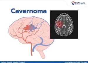
 A Cavernous Malformation (Cavernoma) is an abnormal cluster of tightly packed, thin-walled, slow-flowing blood vessels resembling a raspberry or mulberry, typically located in the brain, brainstem, or spinal cord.
Symptoms
Many cavernomas do not cause symptoms and are found by chance.
When they do cause issues—often appearing between ages 20 and 40—symptoms depend on their size, number, and exact location:
Seizures: Often the most common symptom (especially if located in the cerebral cortex).
Headaches: Sudden or persistent head pain.
Neurological Deficits: Muscle weakness, numbness, or tingling in the face or limbs.
Vision & Balance Issues: Double vision, dizziness, or trouble walking.
Hemorrhage: Minor or major bleeding into surrounding brain tissue, which can mimic a stroke.
Sporadic: Most cases occur randomly as a single lesion with no clear family history.
Familial: About 20% to 30% of cases are inherited in an autosomal dominant pattern linked to specific gene mutations (CCM1/KRIT1, CCM2, or CCM3/PDCD10), often resulting in multiple lesions.
Prior radiation therapy to the brain or spine can also trigger their formation.
Diagnosis
MRI: Magnetic resonance imaging (specifically using susceptibility-weighted or T2-weighted/GRE sequences) is the gold standard, showing a characteristic “popcorn-like” or berry-like appearance.
Genetic Testing: Recommended for individuals with multiple cavernomas or a strong family history.
Management and Treatment
Watchful Waiting: Asymptomatic or stable lesions are frequently monitored over time with routine MRI scans.
Medications: Anti-seizure drugs or pain relievers are used to manage symptoms like epilepsy or headaches.
Surgical Removal: Microsurgery by craniotomy may be recommended if the cavernoma causes drug-resistant seizures, severe or repeated hemorrhages, or grows in an accessible area.