 A Brodmann area is a region of the cerebral cortex.
It defined by its cytoarchitecture, or histological structure and organization of cells.
Brodmann areas were originally defined and numbered on the cytoarchitectural organization of neurons he observed in the cerebral cortex using the Nissl method of cell staining.
Brodmann areas are the most widely known and frequently cited cytoarchitectural organization of the human cortex.
Many of the areas Brodmann defined based solely on their neuronal organization have since been correlated closely to diverse cortical functions.
Brodmann areas 3, 1 and 2 are the primary somatosensory cortex; area 4 is the primary motor cortex; area 17 is the primary visual cortex; and areas 41 and 42 correspond closely to primary auditory cortex,
Brodmann area 25 is the anterior cingulate cortex.
Higher order functions of the association cortical areas are also consistently localized to the same Brodmann areas by neurophysiological, functional imaging, and other methods.
There is consistent localization of Broca’s speech and language area to the left Brodmann areas 44 and 45.
Functional imaging can only identify the approximate localization of brain activations in terms of Brodmann areas since their actual boundaries in any individual brain requires its histological examination.
Different parts of the cerebral cortex are involved in different cognitive and behavioral functions.
The differences manifest in a number of ways: the effects of localized brain damage, regional activity patterns exposed when the brain is examined using functional imaging techniques, connectivity with subcortical areas, and regional differences in the cellular architecture of the cortex.
Most of the cortex, is called the neocortex that as having six layers.
These layers are not apparent in all areas, and even when a layer is present, its thickness and cellular organization may vary.
Brodmann, split the cortex into 52 different areas and assigned each a number.
Many of these Brodmann areas have since been subdivided.
Many brain areas defined by Brodmann have their own complex internal structures.
Many brain areas are organized into topographic maps, where adjoining bits of the cortex correspond to adjoining parts of the body, or of some more abstract entity.
The primary motor cortex, a strip of tissue running along the anterior edge of the central sulcus.
Motor areas innervating each part of the body arise from a distinct zone, with neighboring body parts represented by neighboring zones.
Electrical stimulation of the cortex at any point causes a muscle-contraction in the represented body part.
The somatotopic representation is not evenly distributed: the head, for is represented by a region about three times as large as the zone for the entire back and trunk.
The size of any zone correlates to the precision of motor control and sensory discrimination possible.
The areas for the lips, fingers, and tongue are particularly large, considering the proportional size of their represented body parts.
The maps for visual areas are retinotopic topography of the retina:is uneven: the fovea—the area at the center of the visual field—is greatly overrepresented compared to the periphery.
The visual circuitry in the human cerebral cortex contains several dozen distinct retinotopic maps.
Brodmann area 17, the primary visual cortex is the main recipient of input from the visual part of the thalamus.
Other visual areas farther downstream extract features such as color, motion, and shape.
In auditory areas, the primary map is tonotopic where sounds are parsed according to frequency by subcortical auditory areas.
There are a number of tonotopic cortical maps, each devoted to analyzing sound in a particular way.
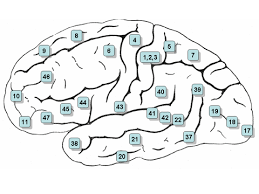
Areas 3, 1 and 2 – Primary somatosensory cortex in the postcentral gyrus (frequently referred to as Areas 3, 1, 2)
Area 4– Primary motor cortex
Area 5 – Superior parietal lobule
Area 6 – Premotor cortex and Supplementary Motor Cortex
Area 7 – Visuo-Motor Coordination
Area 8 – Includes Frontal eye fields
Area 9 – Dorsolateral prefrontal cortex
Area 10 – Anterior prefrontal cortex
Area 11 – Orbitofrontal area
Area 12 – Orbitofrontal area
Area 13 and Area 14* – Insular cortex
Area 15– Anterior Temporal lobe
Area 16 – Insular cortex
Area 17 – Primary visual cortex (V1)
Area 18 – Secondary visual cortex (V2)
Area 19 – Associative visual cortex (V3, V4, V5)
Area 20 – Inferior temporal gyrus
Area 21 – Middle temporal gyrus
Area 22 – Part of the superior temporal gyrus, included in Wernicke’s area
Area 23 – Ventral posterior cingulate cortex
Area 24 – Ventral anterior cingulate cortex.
Area 25 – Subgenual area (part of the Ventromedial prefrontal cortex)
Area 26 – Ectosplenial portion of the retrosplenial region of the cerebral cortex
Area 27 – Presubiculum
Area 28 – Ventral entorhinal cortex
Area 29 – Retrosplenial cortex
Area 30 – Subdivision of retrosplenial cortex
Area 31 – Dorsal Posterior cingulate cortex
Area 32 – Dorsal anterior cingulate cortex
Area 33 – Part of anterior cingulate cortex
Area 34 – Dorsal entorhinal cortex (on the Parahippocampal gyrus)
Area 35 – Part of the perirhinal cortex (in the rhinal sulcus)
Area 36 – Part of the perirhinal cortex (in the rhinal sulcus)
Area 37 – Fusiform gyrus
Area 38 – Temporopolar area (most rostral part of the superior and middle temporal gyri)
Area 39 – Angular gyrus, considered by some to be part of Wernicke’s area
Area 40 – Supramarginal gyrus considered by some to be part of Wernicke’s area
Areas 41 and 42 – Auditory cortex
Area 43 – Primary gustatory cortex
Areas 44 and 45 – Broca’s area, includes the opercular part and triangular part of the inferior frontal gyrus
Area 46 – Dorsolateral prefrontal cortex
Area 47 – Orbital part of inferior frontal gyrus
Area 48 – Retrosubicular area (a small part of the medial surface of the temporal lobe)
Area 49 – Parasubicular area
Area 52 – Parainsular area