1591
1349
Develops from a strip of ectoderm.
Human brain growth is most rapid during the third trimester of pregnancy and continues to be rapid to approximately five years of age.
Continues to develop postnatally with heightened vulnerability into early childhood.
Most neurons are formed by the time of birth, glial cells and myelinization of axons continues for several years.
Brain development is largely completed in utero.
Limited neurogenesis continues throughout childhood and adult life but only in specific regions of the brain: dentate gyrus of the hippocampus and the sub ventricular zone of the striatum.
Parenchyma made up of 80% water.
The average weight of the male brain is 1,336 gms and the female brain is 1,198 gms.
The weight of the brain decreases in advancing age.
The human brain is the fattest organ in the body and may consist of at least 60% fat.
Despite the brain being highly enriched in cholesterol, the blood-brain-barrier prevents influx of lipids and lipoproteins, including cholesterol, from the blood to the brain.
Unlike peripheral tissues, which obtain blood-borne cholesterol secreted by the liver, the brain synthesizes all its cholesterol de novo.
The primary fuel source is glucose, which is neither synthesized nor stored in the brain.
Estimated 86 billion neurons in the brain.
The adult human brain contains about 86 billion neurons, of which 16.3 billion are in the cerebral cortex and 69 billion in the cerebellum.
The brain, which accounts for only 2 percent of total body weight, typically receives 12 percent of blood flow from the heart.
At the beginning of adolescence cellular proliferation of the brain had stopped, and ongoing brain development active through regional myelinization, cortical maturation, neural reconnections, and axonal hypertrophy (Casey BJ et al).
High cognitive processing regulated primarily in the prefrontal region and is dependent on widely distributed subcortical networks between the cortex, basal ganglia and thalamus.
Frontal brain regions are integral for executive functioning, as well as maintenance of functionality with age, and the ability to successfully complete activity of daily living.
Cognitive processes sensitive to genetic, metabolic and hypoxic processes, exposure to toxins, trauma and brain tumors.
The brain has information processing circuits, or systems, and should be viewed not as isolated neurons and regions.
Brain volume does not correlate to intelligence, or intelligence quotient (IQ).
Each of the one hundred billion neurons of the brain has on average 7,000 synaptic connections to other neurons.
Brain disorders reflect problem in information flow across circuits.
The brain has antagonistic systems for promoting and inhibiting movement.
Cerebral cortex composed of specialized regions labeled as Brodmann’s areas.
Cerebral cortex has 6 layers of cells and the relative size of each layers varies among the regions of the brain.
Each layer of the cerebral cortex has specialized cells.
Cerebral cortex divided into the frontal, parietal, temporal, and occipital lobes.
The sylvian fissure divides the temporal lobes from the rest of the cortex.
The central sulcus divides the frontal from the parietal lobe.
The sagital sulcus divides the brain into the left and right hemispheres.
The frontal lobe lies anterior to the central sulcus and is composed of the prefrontal cortex, premotor cortex, primary motor cortex, frontal eye fields and Broca’s area.
Areas related to movement include the primary motor cortex and premotor cortex located anterior to the central gyrus.
The primary motor cortex of the cerebral cortex located closest to the central sulcus and the premotor cortex is located anteriorly in the frontal lobe.
The premotor cortex is related to planning movements and the primary motor cortex executes the movements via the descending motor neurons of the corticospinal tract.
Areas of the motor cortex correspond to the body parts they control, and a lesion of the motion strip leads to spastic paralysis of the contralateral body part corresponding to the damaged site.
Broca’s area 8, the frontal eye fields, control eye movements.
Lesions of the frontal eye fields lead to the eyes drifting down toward the side of the injured area.
Hyperactivity of the frontal eye fields leads to eye drift away from the side of hyperactivity.
The frontal lobe is associated with inhibiting thoughts to promote social norms.
Lesions of the prefrontal cortex leads to disinhibition.
Language production is involved with area 44 (Broca‘s area).
Almost all right-handed people have a left cerebral hemisphere, which is responsible for language.
Lesions in Broca’s area leads to an inability of producing coherent speech, with incomprehensible words difficulty forming words and slow and laborious speech.
Executive functions associated with the prefrontal cortex with concentration, judgment, orientation, abstraction and inhibition of primitive reflexes.
Lesions of the prefrontal cortex can unmask primitive reflexes such as suckling, rooting and grasp reflexes.
The anterior cerebral artery and middle cerebral artery supply the blood to the frontal lobe.
Parietal lobes are located lateral to the sagital sulcus and posterior to the frontal lobes and posterior to the central sulcus.
Parietal lobes are supplied by the middle cerebral artery and the anterior cerebral artery in the region of the homunculus which supplies the leg.
Parietal lobes are the primary sensory cortex.
The primary sensory centers of the parietal lobe are posterior to the central sulcus and the primary motor cortex.
A sensory homunculus represents the anatomical correlation like the motor cortex.
The ascending spinal thalamic and dorsal spinal tracts synapse in the thalamus and project to the primary sensory cortex of the parietal lobe.
The primary sensory cortex of the parietal lobe sends projections to the secondary and associated cortices that integrate the sensory components.
Lesions of the parietal lobe leads to loss of tactile sensation in areas of the body corresponding to the injured areas of the brain.
Parietal lobe determines spatial relationships of right and left.
Lesions of the inferior parietal lobe of the dominant hemisphere associated with Gerstmann’s syndrome, manifesting right and left sided confusion, inability to name and recognize one’s own fingers or others’ fingers, inability to write and read and inability to make arithmetic calculations.
Parietal lobe lesion may cause contralateral hemianopsia and lower quadrantic hemianopsia due to injury of the optic tract passing through the parietal lobe on the way to the occipital lobe.
Unilateral lesions of the parietal lobe may lead to contralateral neglect, or the patient may draw a clock withal the numbers on one side.
Lesions of the dominant parietal hemisphere can cause apraxia, the inability to carry out learned movements.
Bilateral lesions of the parietal lobe can lead to Balint’s syndrome, a type of visual agnosia.
The middle cerebral artery and the anterior cerebral arteries supply the homunculus of the leg.
Injuries occurring in the parietal lobe optic tract as it passes to the occipital lobe can cause contralateral hemianopsia and lower quadrant hemianopsia.
When one looks at an object, the image of that object appears upside down on their retina.
However, your brain automatically corrects for this, allowing you to perceive the object the right side up.
Unilateral lesion may cause contralateral neglect.
Unilateral parietal lesion may cause clock drawing with all numbers of one side.
Apraxia may be a result of a parietal dominant hemisphere lesion.
The occipital lobe is the most posterior part of the brain an it is the primary visual cortex and association visual cortex.
The visual pathway begins at the retina goes to fibers that cross at the optic chiasma then synapses at the lateral genticulate nucleus and goes to the primary visual cortex within the occipital lobe.
Visual signals processed from the primary visual cortex to the visual association cortex in the occipital lobe.
Lesions of the visual association cortex can lead to visual agnosia, the inability to recognize objects one sees.
Lesions in the visual association cortex can lead to alexia without agraphia, the aired inability to read while retaining the ability to write.
The blood supply to the occipital lobe is via the posterior cerebral artery.
Temporal lobes of the bran contain structures vital forhearing, memory and emotion.
Temporal lobe is located lateral to the sylviam fissure and are outpouchings of the cortex.
The major areas of the temporal lobe include the auditory cortex, the hippocampus amygdala and Wernicke’s area.
The primary auditory cortex is located within the sylvian fissure.
Basal ganglia refers to five structures on each side of the brain:caudate nucleus, putamen, globus pallidus, which or three lower nuclear masses underlying the cortical mantle, and the subthalamic nucleus and substantia nigra.
The globus pallidus is divided into a an external and internal segment.
The substantia nigra is divided into a pars compacta and a pars reticulata.
The thalamus and the basal ganglia or interrelated.
The caudate nucleus and putamen are referred to as the striatum.
The putamen and the globus pallidus are sometimes reffered to as the lenticular nucleus.
The major afferent connections to the basal ganglia terminate in the striatum.
The output from of the internal segment of the globus pallidus to the thalamus is inhibitory, while the output from the thalamus to the cerebral cortex is excitatory.
Basal ganglia structures have a high oxygen consumption, and the copper content of the substantia nigra and locus ceruleus is very high.
One of the most complex organs in the human body.
Our brain gives us awareness of ourselves, and of our environment.
All thought, feelings and plans come from the brain.
The brain processes a constant stream of sensory data.
It controls our muscle movements.
Controls the secretions of our glands.
Controls breathing and internal temperature.
Weighing only about 3 pounds.
It consumes as much as 20% of the oxygen and glucose taken in by the body.
The human brain consumes approximately 60% of blood glucose in fasted, sedentary individuals.
Nervous tissue in the brain associated with very high metabolic rate due to the number of processes taking place within the brain at any given time.
It is possible to divide the brain into three main regions based on embryonic development: the forebrain, midbrain and hindbrain.
The forebrain (or prosencephalon) is made up of our cerebrum, thalamus, hypothalamus and pineal gland.
The cerebral area is noted as the telencephalon and the term diencephalon to refers to the area where our thalamus, hypothalamus and pineal gland reside.
The midbrain, or mesencephalon, located near the very center of the brain between the interbrain and the hindbrain, is composed of a portion of the brainstem.
The hindbrain, or rhombencephalon, consists of the remaining brainstem as well as our cerebellum and pons.
Brain cells can be broken into two groups: neurons and neuroglia.
Neurons perform all of the communication and processing within the brain.
Sensory neurons entering the brain from the peripheral nervous system deliver information about the condition of the body and its surroundings.
Most of the neurons in the brain’s gray matter are interneurons, which are responsible for integrating and processing information delivered to the brain by sensory neurons.
Brain maturation process throughout the adolescent and young adult years.
Gray matter normally decreases in volume during brain maturation due to neuronal pruning, and white matter increases with myelinization.
In the pre-teens less frequently used neurons are pruned in order to build complex networks for the decision-making of adulthood.
The limbic system that exists below and posterior to the cortex undergoes maturation prior to the gray matter of the prefrontal cortex, that is responsible for logical thought and impulse regulation
Interneurons send signals to motor neurons, carrying signals to muscles and glands.
Neuroglia, or glial cells, the helper cells of the brain support and protect neurons.
In the brain there are four types of glial cells: astrocytes, oligodendrocytes, microglia, and ependymal cells.
Astrocytes protect neurons by filtering nutrients out of the blood and preventing chemicals and pathogens from leaving the capillaries of the brain.
Oligodendrocytes wrap the axons of neurons in the brain to produce the insulation known as myelin.
Myelinated axons transmit nerve signals much faster than unmyelinated axons.
Oligodendrocytes accelerate the communication speed of the brain.
Microglia act like white blood cells by attacking and destroying pathogens that invade the brain.
Ependymal cells line the capillaries of the choroid plexuses and filter blood plasma to produce cerebrospinal fluid.
The tissue of the brain can be broken down into two major classes: gray matter and white matter.
Gray matter is made of mostly unmyelinated neurons, most of which are interneurons, and is the areas of nerve connections and processing.
White matter is made of mostly myelinated neurons that connect the regions of gray matter to each other and to the rest of the body.
Myelinated neurons transmit nerve signals much faster than unmyelinated axons do, and act as the information highway of the brain to speed the connections between distant parts of the brain and body.
The brainstem is the most inferior portion of our brain, and connects the brain to the spinal cord.
Many basic survival functions of the brain are controlled by the brainstem.
The brainstem is made of three regions: the medulla oblongata, the pons, and the midbrain.
The reticular formation is found in all three regions of the brainstem.
The reticular formation controls muscle tone in the body and acts as the switch between consciousness and sleep in the brain.
The medulla oblongata is a cylindrical-like mass of nervous tissue that connects to the spinal cord on its inferior border and to the pons on its superior border.
The medulla oblongata contains mostly white matter that carries nerve signals ascending into the brain and descending into the spinal cord.
Areas of gray matter in the medulla oblongata process involuntary body functions related to homeostasis.
The medulla cardiovascular center monitors blood pressure and oxygen levels and regulates heart rate to provide sufficient oxygen supplies to the body.
The medulla controls the rate of breathing to provide oxygen to the body.
The medulla oblongata gray matter coordinates the vomiting, sneezing, coughing, and swallowing reflexes.
The pons region of the brainstem is found superior to the medulla oblongata, inferior to the midbrain, and anterior to the cerebellum.
The pons and the cerebellum forms the metencephalon.
The pons acts as the bridge for nerve signals traveling to and from the cerebellum and carries signals between the superior regions of the brain and the medulla and spinal cord.
The cerebellum is a hemispherical region of the brain located posterior to the brainstem and inferior to the cerebrum.
The outer layer of the cerebellum is known as the cerebellar cortex.
The cerebeller cortex is made of tightly folded gray matter that provides the processing power of the cerebellum.
Deep to the cerebellar cortex is a tree-shaped layer of white matter called the arbor vitae.
The arbor vitae connects the processing regions of cerebellar cortex to the rest of the brain and body.
The cerebellum helps to control motor functions such as balance, posture, and coordination of complex muscle activities.
The cerebellum receives sensory inputs from the muscles and joints of the body and uses this information to keep the body balanced and to maintain posture.
The cerebellum also controls the timing and complexity of motor actions such as walking, writing, and speech.
The midbrain is the most superior region of the brainstem.
The midbrain is also known as the mesencephalon,
The midbrain is found between the pons and the diencephalon.
It is subdivided into 2 main regions: the tectum and the cerebral peduncles.
The tectum is the posterior region of the midbrain.
The tectum contains relays for reflexes that involve auditory and visual information, the pupillary reflex, eye accommodation reflex, and startle reflex.
The cerebral peduncles form the anterior region of the midbrain, and contains many nerve tracts and the substantia nigra.
Nerve tracts pass through the cerebral peduncles and connect regions of the cerebrum and thalamus to the spinal cord and lower brainstem.
The substantia nigra contains dark melanin-containing neurons in a region of the brain that is involved in the inhibition of movement.
Sibstantia nigra deterioration leads to a loss of motor control known as occurs in Parkinson’s disease.
Superior and anterior to the midbrain is the diencephalon region, with the thalamus, hypothalamus, and pineal glands making up its major regions.
The thalamus consists of a pair of oval masses of gray matter inferior to the lateral ventricles and surrounds the third ventricle.
The peripheral nervous system sensory neurons form relays with neurons in the thalamus that continue on to the cerebral cortex.
The thalamus routes sensory inputs to the correct regions of the cerebral cortex.
The thalamus routes sensory information into processing and memory centers of the cerebrum aiding learning.
The hypothalamus is located inferior to the thalamus and superior to the pituitary gland.
The hypothalamus controls body temperature, hunger, thirst, blood pressure, heart rate, and the production of hormones.
The hypothalamus responds to the body’s sensory receptors reflecting alterations by sending signals to glands, smooth muscles, and the heart to counteract these changes.
The hypothalamus controls the pituitary gland by producing hormones.
Oxytocin and antidiuretic hormone, are produced in the hypothalamus and stored in the posterior pituitary gland.
Other releasing and inhibiting hormones, are secreted into the blood to stimulate or inhibit hormone production in the anterior pituitary gland.
The pineal gland is a small gland located posterior to the thalamus.
The pineal gland produces the hormone melatonin.
Cerebrum is the largest region of the human brain.
The cerebrum controls higher brain functions such as language, logic, reasoning, and creativity, surrounding the diencephalon and is located superior to the cerebellum and brainstem.
The longitudinal fissure runs midsagittally down the center of the cerebrum, dividing the cerebrum into the left and right hemispheres.
Each cerebral hemisphere is divided into 4 lobes: frontal, parietal, temporal, and occipital.
The lobes of the cerebrum are named for the skull bones that cover them.
The surface of the cerebrum, the cerebral cortex, is a convoluted layer of gray matter
The bulges of cortex are called gyri.
Indentations of the cortex are called sulci
Cerebrum processing mainly takes place within the cerebral cortex.
Deep to the cerebral cortex is a layer of cerebral white matter.
White matter contains the connections between the regions of the cerebrum as well as between the cerebrum and the rest of the body.
The corpus callosum, a band of white matter, connects the left and right hemispheres of the cerebrum and allows the hemispheres to communicate with each other.
Deep within the cerebral white matter are regions of gray matter that make up the basal nuclei and the limbic system.
The basal nuclei, including the globus pallidus, striatum, and subthalamic nucleus, work together with the substantia nigra of the midbrain to regulate and control muscle movements.
The basal nuclei, regions help to control muscle tone, posture, and subconscious skeletal muscle.
The limbic system, a group of deep gray matter regions, including the hippocampus and amygdala, are involved in memory, survival, and emotions.
The limbic system helps the body to react to emergency and highly emotional situations with fast, almost involuntary actions.
The brain is protected by the meninges.
The meninges are 3 layers of tissue, surrounding and protecting the brain and spinal cord.
The dura mater forms the outermost layer of the meninges.
The dura mater contains irregular connective tissue made of tough collagen fibers.
It is these collagen fibers that gives the dura mater its strength.
The arachnoid mater is found lining the inside of the dura mater.
The arachnoid mater is thinner and more delicate than the dura mater.
The arachnoid mater contains many fibers that connect the dura mater and pia mater.
Beneath the arachnoid mater is a fluid-filled region known as the subarachnoid space.
The pia mater the innermost of the meningeal layers, rests directly on the surface of the brain and spinal cord.
The pia mater many provides nutrients and oxygen to the nervous tissue of the brain.
The pia mater helps to regulate the flow of materials from the bloodstream and cerebrospinal fluid into nervous tissue.
Cerebrospinal fluid (CSF) surrounds the brain and spinal cord.
The brain and spinal cord float within the CSF.
CSF fills the subarachnoid space and exerts pressure on the outside of the brain and spinal cord, and acts as a stabilizer and shock absorber as they float within the hollow spaces of the skull and vertebrae.
Inside of the brain, CSF-filled cavities called ventricles expand under the pressure of CSF to lift and inflate the soft brain tissue.
Cerebrospinal fluid is produced in the brain by capillaries lined with ependymal cells known as choroid plexuses.
Blood plasma passing through the capillaries is filtered by the ependymal cells and released into the subarachnoid space as CSF.
The CSF contains glucose, oxygen, and ions, which it helps to distribute throughout the nervous tissue.
CSF also transports waste products away from nervous tissues.
After circulating around the brain and spinal cord, CSF enters the arachnoid villi where it is reabsorbed into the bloodstream.
The brain consists of four fluid compartments: cerebrospinal fluid (CSF), interstitial fluid, intracellular fluid, and the blood vasculature.
The fluid compartments in the brain consist of intracellular fluid (ICF) (60-68%), interstitial fluid (ISF) (or extracellular fluid) (12-20%), blood (10%) and the cerebrospinal fluid (CSF) (10%).
The blood is kept separate from the brain parenchyma and CSF by the blood-brain and blood-CSF barriers, respectively.
These barriers are essential in maintaining the extracellular environment of the brain because they regulate the ionic and biochemical composition of the different fluid compartments.
Arachnoid villi of the arachnoid mater pass through the dura mater and into the superior sagittal sinus.
The superior sagittal sinus is a vein that runs through the longitudinal fissure of the brain.
The superior sagittal sinus vein carries blood and cerebrospinal fluid from the brain back to the heart.
Blood must be constantly delivered to the brain to maintain proper brain function.
Any interruption in the delivery of blood to the brain leads very quickly to dizziness, disorientation, and eventually unconsciousness.
It receives information about the body’s condition and surroundings from all of the sensory receptors in the body , creating perception of the body’s internal and external conditions.
The sensory data received but the brain include autonomic and somatic information.
The autonomic sensory information tells the brain about the condition of the body including body temperature, heart rate, and blood pressure: subconscious information.
The somatic sensory information to the brain is awareness of touch, sight, sound, and hearing, which are all examples of somatic conscious senses.
The brain directly controls almost all movement in the body, as the motor area of the cerebral cortex sends signals to the skeletal muscles to produce all voluntary movements.
Subconsciously, the basal nuclei of the cerebrum and gray matter in the brainstem help to control these movements and prevent extraneous motions, while the cerebellum helps with the timing and coordination of these movements during complex motions.
The autonomic regions of the brain stimulate the motor output to smooth muscle tissue, cardiac muscle tissue, and glands
The association areas of the brain process and analyze sensory information, to develop plans of action that are sent to the brain’s motor regions in order to produce a change in the body through muscles or glands.
This association area process also creates our thoughts, plans, and personality.
The brain stores many different types of information.
The brain receives information from the senses and it develops information through thinking in the association areas.
Information in the brain is stored in a few ways depending on its source and how long it is needed.
Short-term memory keeps track of the tasks in which the brain is currently engaged.
Short-term memory probably consists of a group of neurons that stimulate each other in a loop to keep data in the brain’s memory.
New information replaces the old information in short-term memory within a few seconds or minutes, unless the information gets moved to long-term memory.
Long-term memory is stored in the hippocampus of the brain.
The hippocampus transfers information from short-term memory to memory-storage regions of the brain, particularly in the cerebral cortex of the temporal lobes.
Memory related to motor skills is stored by the cerebellum and basal nuclei.
The brain is the body’s control center maintaining the homeostasis of many diverse functions such as breathing, heart rate, body temperature, and hunger.
The brainstem and the hypothalamus are the brain structures most concerned with the body’s homeostasis.
The medulla oblongata of the brain stem contains the cardiovascular center that monitors the levels of dissolved carbon dioxide and oxygen in the blood, along with blood pressure.
The cardiovascular center adjusts the heart rate and blood vessel dilation to maintain healthy levels of dissolved gases in the blood and to maintain a healthy blood pressure.
The medulla rhythmicity center monitors oxygen and carbon dioxide levels in the blood and adjusts the rate of breathing to keep these levels in balance.
The homeostasis of body temperature, blood pressure, sleep, thirst, and hunger are controlled by the hypothalamus.
Autonomic sensory receptors for temperature, pressure, and chemicals feed into the hypothalamus, where it is processed and sends the output to autonomic effectors in the body such as sweat glands, the heart, and the kidneys.
The hypothalamus maintains the body’s 24 hour circadian clock.
When it is time for sleep as indicated by the circadian clock has arrived, it sends signals to the reticular activating system of the brainstem to reduce its stimulation of the cerebral cortex.
The reduction in the stimulation of the cerebral cortex leads to a sleepiness and eventually to sleep.
During sleep state, the brain stops maintaining consciousness, reduces some sensitivity to sensory input, relaxes skeletal muscles, and completes administrative functions, such as storage of memory, dreaming, and development of nervous tissue.
Sleephas two main stages of sleep: rapid eye movement (REM) and non-rapid eye movement (NREM).
During REM sleep, the muscles of the body becomes paralyzed while the eyes move back and forth quickly.
Dreaming is common during REM sleep
Some memories are stored during REM phase.
NREM sleep is a period of slow eye movement or no eye movement, culminating in a deep sleep of low brain electrical activity.
Dreaming during NREM sleep is rare.
Memories are still processed and stored during NREM time.
A reflex refers to fast, involuntary reaction to a form of internal or external stimulus.
The brain integrates many reflexes including the pupillary light reflex, coughing, and sneezing.
Reflexes frequently protect the body from harm.
All reflexes occur by bypassing the control centers of the cerebral cortex and integrating in the lower regions of the brain such as the midbrain or limbic system.
In Wilson’s disease, in which plasma levels of copper-binding protein ceruloplasmin is low chronic copper intoxication occurs and degeneration of the lenticular nucleus takes place.
The brain’s vasculature has unique features that distinguishes it from the vasculature in the rest of the body.
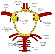
The arterial cerebral circulation consists of an anterior cerebral circulation and posterior cerebral circulation supplied by the internal carotid arteries and the vertebral arteries, respectively.
The anterior circulation, which includes the middle and anterior cerebral arteries, communicates with the posterior circulation, the basilar artery and posterior cerebral arteries, via anterior and posterior communicating arteries at the Circle of Willis.
From the Circle of Willis, the anterior circulation perfuse the younger parts of the brain including the neocortex of the cerebral hemispheres, while the posterior circulation supplies the brainstem and cerebellum.
At the cortical surface, cerebral arteries extend into pial arteries running through the CSF-containing subarachnoid space and the subpial space.
As pial arteries dive down into the brain parenchyma they transition into penetrating arterioles and create a perivascular space, known as the Virchow-Robin space.
The Virchow-Robin spaces are filled with CSF and bordered by a leptomeningeal cell layer on both the inner wall facing the vessel and on the outer wall facing perivascular astrocytic endfeet.
One reply on “Brain”
[…] The large mass of gray matter in the dorsal part of the diencephalon of the brain. […]