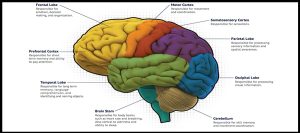
 Brain mapping is specifically defined, as the study of the anatomy and function of the brain and spinal cord through the use of imaging, immunohistochemistry, molecular & optogenetics, stem cell and cellular biology, engineering, neurophysiology and nanotechnology.
All neuroimaging is considered part of brain mapping.
It is considered a higher form of neuroimaging.
It produces brain images supplemented by the result of additional, imaging or non-imaging, data processing or analysis, such as maps projecting behavior onto brain regions.
A connectogram is a map that depicts cortical regions around a circle, organized by lobes.
Concentric circles illustrate the connections between cortical regions, weighted by and strength of connection.?
Connectomes incorporate individual neural connections in the brain and are often presented as wiring diagrams.
Brain mapping techniques rely on the development and refinement of image acquisition, representation, analysis, visualization and interpretation techniques.
Functional and structural neuroimaging are the basis of of brain mapping.
Tecnniques used for brain mapping include:
functional magnetic resonance imaging (fMRI), diffusion MRI (dMRI), magnetoencephalography (MEG), electroencephalography (EEG), positron emission tomography (PET), Near-infrared spectroscopy (NIRS).
Brains may be mapped to study memory, learning, aging, and drug effects in various populations such as people with schizophrenia, autism, and clinical depression.
