
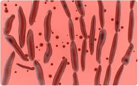
 Acute neurological process with neuroparalytic due to a neurotoxin produced by Clostridium botulinum.
Neurotoxin binds irreversibly to presynaptic membranes of peripheral neuromuscular and autonomic nerve junctions.
C botulinum is an anaerobic gram positive rod that survives in the soil and marine sediment by spore formation.
Under anaerobic conditions C botulinum germinates and releases an exotoxin.
The botulinum organism stains gram positively early , but may be gram negative after 18 hours of incubation, making diagnosis more complicated.
The binding of the neurotoxin blocks acetylcholine release with weakness, paralysis and possible respiratory arrest.
The toxin mediated infection causes blockage of the transmission of acetylcholine across cholinergic synapses of the peripheral nervous system resulting in descending, flaccid, symmetric pattern of paralysis, with prominent bulbar palsy.
Presents as a symmetrical, descending motor paralysis, but symptoms vary in severity from sub clinical to fatal, and the rate of progression depends on the neurotoxin serotype and exposure quantity.
Symptoms usually begin two hours to eight days after exposure of the neurotoxin with the peak at 48 hours.
Associated with loss of ocular accommodation, diplopia, blurred vision, dysphonia, dysarthria, and facial weakness.
The sensorium is unaffected.
Symmetrical cranial palsies are often initial symptoms, manifesting as difficulties with vision or eye movement or bulbar weakness with dysphasia, dysarthria, and dysphonia.
General, weakness, vomiting, nausea, dry mouth, abdominal pain are common.
1/3 of cases have respiratory distress.
Without treatment symptoms can progress to symmetric limb motor weakness, and neuromuscular respiratory failure, resulting from diaphragmatic weakness.
Cranial nerve palsies can contribute to respiratory failure, resulting in pharyngeal collapse, and airway compromise.
Differential diagnosis includes Guillain-Barre syndrome, LEMS, myasthenia gravis, Bickerstaff’s brainstem encephalitis, viral encephalitis, a severe myopathy, and periodic paralytic shellfish poisoning
3 presentations of the disease are infant type, foodborne and wound type.
Infant type related to ingestion of C botulinum spores that germinate in the intestine.
Infant type usually results in full recovery with supportive care and a mortality rate of less than 1%.
In the infant type the spores come from bee honey or the environment.
Foodborne botulism related mainly to improperly canned foods or contaminated home prepared foods.
Wounds contaminated with toxin producing C botulinum associated with wound botulism.
Foodborne disease and wound disease occur primarily in adults, and is rare.
Toxin production can occur in foods with low acidity, a low salt content, or a low sugar content that are contaminated with viable C. botulinum spores and have been left at ambient temperatures in low oxygen conditions.
There are eight antigen specific C botulinum toxins: A, B, C (alpha), C (beta), D, E, F and G.
Types A, B,E, cause disease, and F rarely does so.
Type A and B toxins are the most potent.
Toxin A most commonly involved with foodborne outbreaks, followed by B and E.
In 15% of cases no toxin is identified.
Type G botulinum toxin associated with sudden deafness.
Botulinum toxin is produced by Clostridium botulinum a spore forming anaerobic gram-positive bacterium found in soil, dust, and aquatic sediment.
There are 70 types of botulinum neurotoxin types A through G all of which caused paralysis by the irreversibly inhibiting acetylcholine release in the neuromuscular junction of voluntary motor and autonomic morons.
Diagnosis: the diagnosis is confirmed by the detection of neurotoxin in serum with the use of a combination of bioassays and molecular tests.
The sensitivity of the tests is dependent on the timing of the sample collection relative to the onset of the symptoms and the administration of antitoxin, as well as thecause of the poisoning and absorption and metabolism kinetics of the neurotoxin.
EMG and nerve conduction studies can help as they often show small compound muscle action potential and incremental increase increases in amplitude with frequency repetitive nerve stimulation.
Treatment: includes the administration of equine derived heptavalent antitoxin which inactivates all the antitoxin serotypes, that does not involve an infant.
The antitoxin can arrest disease progression, but it does not reverse established paralysis:reversal of paralysis requires the development of new nerve terminals.
The administration of the antitoxin more than 48 hours after the onset of symptoms is associated with longer hospital, stays and decrease survival.
Antitoxin adverse reactions are uncommon and anaphylaxis occurs in approximately 1.4% of patients.
Supportive care includes mechanical ventilation.
Mortality remains high at 10% even among patients receiving the antitoxin due to respiratory failure or sequelae of paralysis.
Patients who survive often recover lung muscle strength within one year, but fatigue exercise intolerance general weakness and dry mouth can persist.