 Acute-phase proteins (APPs) are a class of proteins whose concentrations in blood plasma either increase or decrease in response to inflammation.
The terms acute-phase protein and acute-phase reactant (APR) are often used synonymously, although some APRs are polypeptides rather than proteins.
This response is called the acute-phase reaction.
The acute-phase reaction characteristically involves fever, acceleration of peripheral leukocytes, circulating neutrophils and their precursors.
An acute-phase response is characterized by decreased production of albumin by hepatocytes, reorientation of iron metabolism, and hormonal changes.
Acute-phase proteins serve as fundamental diagnostic tools that have applications in patients with a range of conditions, including infection, cardiovascular illness, cancer, neurodegeneration, and dysmetabolism.
Acute-phase proteins such as C-reactive protein, fibrinogen and its degradation product d-dimer, and ferritin serve as tools in the day-to-day management of illnesses and as prognostic indicators.
A fundamental function of the acute-phase response is to amplify antimicrobial resistance and tissue repair, with many of the acute-phase proteins serving as key components of humoral innate immunity.
The terms acute-phase protein and acute-phase reactant (APR) are often used synonymously, although some APRs are polypeptides rather than proteins.
In response to injury, local inflammatory cells, neutrophil granulocytes and macrophages, secrete cytokines into the bloodstream, most notable of which are the interleukins IL1, and IL6, and TNF-α.
The liver responds to injury/inflammation by producing many acute-phase reactants.
Increased acute-phase proteins from the liver may also contribute to the promotion of sepsis.
With injury/inflammation the production of a number of other proteins are reduced; these proteins are, therefore, referred to as “negative” acute-phase reactants.
TNF-α, IL-1β and IFN-γ are important for the expression of inflammatory mediators such as prostaglandins and leukotrienes, and they also cause the production of platelet-activating factor and IL-6.
Proinflammatory cytokines stimulate Kupffer cells, produce IL-6 in the liver and present it to the hepatocytes.
IL-6 is the major mediator for the hepatocytic secretion of APPs.
Synthesis of APP can also be regulated indirectly by cortisol.
Cortisol can enhance expression of IL-6 receptors in liver cells and induce IL-6-mediated production of APPs.
APPs have different physiological functions within the immune system: Some act to destroy or inhibit growth of microbes-C-reactive protein, mannose-binding protein, complement factors, ferritin, ceruloplasmin, serum amyloid A and haptoglobin.
Others give negative feedback on the inflammatory response-serpins.
Alpha 2-macroglobulin and coagulation factors affect coagulation, mainly stimulating it, and may limit infection by trapping pathogens in local blood clots.
Some coagulation system products contribute to the innate immune system by their ability to increase vascular permeability and act as chemotactic agents for phagocytic cells.
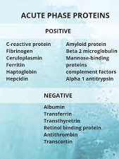
Positive acute-phase proteins:
C-reactive protein-Opsonin on microbes
Serum amyloid P component Opsonin
Serum amyloid A-Recruitment of immune cells to inflammatory sites,
Induction of enzymes that degrade extracellular matrix.
Complement factors Opsonization, lysis and clumping of target cells. Chemotaxis
Mannan-binding lectin pathway of complement activation
Fibrinogen, prothrombin, factor VIII,
von Willebrand factor Coagulation factors, trapping invading microbes in blood clots.
Plasminogen activator inhibitor-1 (PAI-1)-Prevents the degradation of blood clots by inhibiting tissue Plasminogen Activator (tPA)
Alpha 2-macroglobulin
Inhibitor of coagulation by inhibiting thrombin.
Inhibitor of fibrinolysis by inhibiting plasmin
Ferritin Binding iron, inhibiting microbe iron uptake
Hepcidin Stimulates the internalization of ferroportin, preventing release of iron bound by ferritin within intestinal enterocytes and macrophages.
Ceruloplasmin Oxidizes iron, facilitating for ferritin
Haptoglobin Binds hemoglobin, inhibiting microbe iron uptake and prevents kidney damage
Orosomucoid
(Alpha-1-acid glycoprotein, AGP) Steroid carrier
Alpha 1-antitrypsin Serpin, downregulates inflammation
Alpha 1-antichymotrypsin Serpin, downregulates inflammation
Negative
Negative acute-phase proteins decrease in inflammation:
Albumin, transferrin, transthyretin, retinol-binding protein, antithrombin, transcortin.
The decrease of such proteins may be used as markers of inflammation.
The decreased synthesis of such proteins is generally to save amino acids for producing positive acute-phase proteins more efficiently.
The production of C3 increases in the liver with inflammation, but the plasma concentration often lowers because of an increased turn-over: therefore, it is often seen as a negative acute-phase protein.
C-reactive protein, is a useful marker of inflammation that correlates with the erythrocyte sedimentation rate (ESR), however not always directly.
The ESR being largely dependent on the elevation of fibrinogen, an acute phase reactant with a half-life of approximately one week.
The ESR remains higher for longer despite the removal of the inflammatory stimuli.
In contrast, C-reactive protein has a half-life of 6–8 hours that rises rapidly and can quickly return to within the normal range if treatment is employed.