Zinc is an essential trace nutrient required for the proper function of more than 100 enzymes.
Zinc plays a crucial role in nucleic acid metabolism.
An autosomal recessive disorder postulated to occur as a result of mutations in the SLC39A4 gene located on band 8q24.3.
The SLC39A4 gene encodes a transmembrane protein that is part of the zinc/iron-regulated transporter–like protein (ZIP) family and is required for zinc uptake.
Zinc deficiency is likely to occur as a result of a mutation in a zinc transport protein encoded by the SLC39A4 gene and perhaps alteration in a zinc transport ligand.
The zinc/iron-regulated transporter–like protein (ZIP) is highly expressed in the enterocytes in the duodenum and jejunum.
Individuals with this disorder have a decreased ability to absorb zinc from dietary sources.
The absence of a binding ligand needed to transport zinc may contribute to zinc malabsorption.
Differentiating acquired zinc deficiency disorders from acrodermatitis enteropathica have similar clinical presentations.
Can only be accurately diagnosed after attempts to remove zinc supplementation have failed.
Transient acquired zinc deficiencies can occur in premature infants secondary to their greater physiological demand for zinc and lower body stores.
Zinc deficiency can present in full-term breastfed infants as a result of low maternal serum zinc levels or a defect in mammary zinc secretion.
Infants are irritable, with slowing or cessation of growth and development.
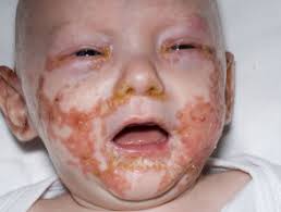
Skin lesions are erythematous, dry, and scaly patches and plaques that may evolve into crusted, vesiculobullous, erosive, psoriasiform, and pustular lesions.
Lesions are predominantly distributed in a perioral and in an acral pattern.
Lesions may become secondarily infected with Staphylococcus aureus or Candida albicans.
Mucosal lesions include angular cheilitis, glossitis, conjunctivitis, blepharitis, punctate keratopathy, and photophobia.
Typical nail findings include paronychia and nail dystrophy.
Loss of scalp hair, eyebrows, and eyelashes occur.
Differential Diagnoses include: Acquired zinc deficiency, Atopic Dermatitis, Candidiasis, Mucosal Epidermolysis Bullosa and Seborrheic Dermatitis.
Plasma zinc levels should be evaluated and concentrations of less than 50 mcg/dL are suggestive, but not diagnostic.
Hair, saliva, or urine zinc levels can be obtained and are additional corroborative tests.
Alkaline phosphatase a zinc-dependent enzyme results in reduced serum levels in the context of normal zinc levels can indicate a zinc deficiency.
Early skin biopsy lesions show confluent parakeratosis associated with a reduced granular layer.
Exocytosis of neutrophils into the epidermis occurs, as does intracellular edema of the upper third of the epidermis.
Subcorneal and intraepidermal clefts may develop from ballooning and reticular degeneration, with necrosis of the keratinocytes.
Degeneration of keratinocytes with formation of multiple cytoplasmic vacuoles and fingerlike protrusions.
The basal lamina is well preserved.
Treatment requires lifelong zinc supplementation.
Typically, 1-3 mg/kg of zinc gluconate or sulfate is given daily.
Clinical improvement occurs within days to weeks of initiating treatment.
To monitor serum zinc levels and alkaline phosphatase values every 3-6 months.
Acrodermatitis enteropathica exacerbation during pregnancy or stress may require an increased therapy.
Removing the scale from the skin surface, followed by adding petrolatum to eroded skin lesions, may enhance reepithelialization.
No special diet is required for treatment as long as zinc supplementation is continued.
Foods with increased levels of zinc include: beef, crab, fowl, oysters, and pork.
Zinc content in foods is directly related to protein content.
High-dose zinc supplementation can cause gastric upset and can adversely affect copper metabolism.
Complications of AE are secondary wound infections, and ultimately death if untreated.
With zinc treatment the response rate is 100%.

