
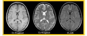
 T2-weighted imaging is an MRI sequence that provides high-resolution anatomical detail of the prostate, allowing clear visualization of the zonal anatomy and identification of suspicious lesions based on differences in tissue signal intensity.
In prostate cancer evaluation, T2-weighted images are essential for detecting and localizing tumors, especially in the peripheral zone, and for assessing features such as extracapsular extension and seminal vesicle invasion.
In biparametric MRI (bpMRI), T2-weighted imaging is one of the two core sequences (along with diffusion-weighted imaging) used to detect and characterize prostate lesions without the need for intravenous contrast.
Radiologists use T2-weighted images to identify areas of low signal intensity that may represent cancer, and these findings are integrated with diffusion-weighted imaging to assign risk scores (e.g., PI-RADS) and guide biopsy decisions.
In multiparametric MRI (mpMRI), T2-weighted imaging is combined with diffusion-weighted and dynamic contrast-enhanced (DCE) imaging.
While T2-weighted imaging provides the anatomical framework, DWI adds functional information about tissue cellularity, and DCE assesses vascularity.
The combination improves lesion characterization and staging, but T2-weighted imaging remains the foundation for anatomical assessment and is critical for both bpMRI and mpMRI protocols.
T2-weighted imaging is the anatomical backbone of both bpMRI and mpMRI for prostate cancer, enabling lesion detection, localization, and staging, and is indispensable for accurate risk stratification and biopsy targeting.
T2 weighted imaging is a magnetic resonance imaging (MRI) technique that highlights differences in the T2 relaxation times of various tissues, primarily representing the decay of transverse magnetization after an RF pulse.
In T2 weighted images, fluids such as cerebrospinal fluid and edema appear bright, making it valuable for identifying pathological conditions like inflammation, edema, and some types of tumors or lesions.
T2 decay refers to the loss of transverse magnetization over time after a 90° RF pulse.
The time constant T2 is defined as the time it takes for the signal from transverse magnetization to decay to about 37% of its initial value.
T2 weighted images are best achieved by using a long echo time (TE) and a relatively long repetition time (TR), allowing differences in T2 times to dominate the image contrast.
Fluids (e.g., water, cerebrospinal fluid) appear bright, as they have long T2 relaxation times. Fat and many normal tissues have shorter T2 times and appear darker in comparison to fluids. Dense tissues (e.g., bone, tendons, areas of calcification) and air appear dark.
Clinical Applications of T2: Commonly used for detecting lesions, edema, and inflammatory processes.
Helpful in neuroimaging, musculoskeletal imaging, and abdominal imaging, since it makes fluid and pathological changes stand out.
T2 weighted imaging is especially valuable in brain MRI to visualize areas affected by stroke, tumors, or demyelinating diseases because these pathologic changes generally increase tissue water content, resulting in brighter areas.
Comparison with T1 Weighted Imaging T1 weighted images emphasize differences in T1 relaxation times and are better for visualizing normal anatomy, where fat appears bright.
T2 emphasizes disease and pathology, as most disease processes involve increased water content, which appears bright on T2.