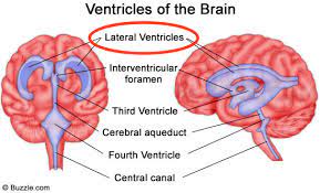
The ventricular system is a set of four interconnected cavities known as cerebral ventricles in the brain.
Within each ventricle is a region of choroid plexus which produces the circulating cerebrospinal fluid (CSF).
The ventricular system is continuous with the central canal of the spinal cord from the fourth ventricle, allowing for the flow of CSF to circulate.
The ventricular system accounts for the production and circulation of cerebrospinal fluid.
All of the ventricular system and the central canal of the spinal cord are lined with ependyma, a specialized form of epithelium connected by tight junctions that make up the blood–cerebrospinal fluid barrier.
The ventricular system is comprised of four ventricles:
lateral ventricles right and left, one for each hemisphere
third ventricle
fourth ventricle
There are several foramina, openings channels, that connect the ventricles.
The interventricular foramina, also called the foramina of Monro, connect the lateral ventricles to the third ventricle through which the cerebrospinal fluid can flow.
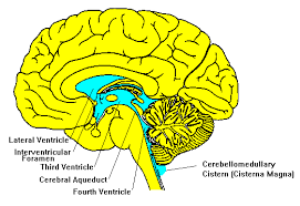
The four cavities of the human brain are called ventricles.
The two largest are the lateral ventricles in the cerebrum.
The third ventricle is in the diencephalon of the forebrain between the right and left thalamus, and the fourth ventricle is located at the back of the pons and upper half of the medulla oblongata of the hindbrain.
The ventricles are concerned with the production and circulation of cerebrospinal fluid.
The ventricular system are embryologically derived from the neural canal, the centre of the neural tube.
The cerebrospinal fluid passes out through arachnoid villi into the venous sinuses of the skull.
The ventricles are filled with cerebrospinal fluid (CSF) which bathes and cushions the brain and spinal cord within their bony confines.
CSF is produced by modified ependymal cells of the choroid plexus found in all components of the ventricular system except for the cerebral aqueduct and the posterior and anterior horns of the lateral ventricles.
CSF flows from the lateral ventricles via the interventricular foramina into the third ventricle, and then the fourth ventricle via the cerebral aqueduct in the midbrain.
From the fourth ventricle CSF can pass into the central canal of the spinal cord or into the subarachnoid cisterns via three small foramina: the central median aperture and the two lateral apertures.
Traditionally it has been held that the majority of CSF is produced by the choroid plexus, circulates through the ventricles, the cisterns, and the subarachnoid space to be absorbed into the blood by the arachnoid villi.
The fluid around the superior sagittal sinus is reabsorbed via the arachnoid granulations/villi into the venous sinuses, after which it passes through the jugular vein and major venous system.
CSF within the spinal cord can flow all the way down to the lumbar cistern at the end of the cord around the cauda equina.
The cerebral aqueduct between the third and fourth ventricles is very small, as are the foramina, which means that they can be easily blocked.
The brain and spinal cord are covered by the meninges.
The meninges are the three protective membranes of the tough dura mater, the arachnoid mater and the pia mater.
CSF in the skull and spine provides protection and buoyancy, and is found in the subarachnoid space between the pia mater and the arachnoid mater: protects the brain from jolts and knocks to the head.
The CSF that is produced in the ventricular system is also necessary for chemical stability, and the provision of nutrients needed by the brain.
The CSF helps to protect the brain from jolts and knocks to the head and also provides buoyancy and support to the brain against gravity:
the brain floats in neutral buoyancy, suspended in the CSF, allowing the brain to grow in size and weight without resting on the floor of the cranium, which would destroy nervous tissue.
The narrowness of the cerebral aqueduct and foramina means that they can become blocked.
Blood that accumulates following a hemorrhagic stroke, can block the ventricular pathways.
CSF is continually produced by the choroid plexus within the ventricles, and a blockage of outflow leads to increasingly high pressure in the lateral ventricles leading to hydrocephalus.
An endoscopic third ventriculostomy is a surgical procedure for the treatment of hydrocephalus.
With a third ventriculostomy an opening is created in the floor of the third ventricle using an endoscope placed within the ventricular system through a burr hole.
The cerebrospinal fluid can then flow directly to the basal cisterns, thereby bypassing any obstruction.
The surgical procedure to make an entry hole to access any of the ventricles is called a ventriculostomy, and is done to drain accumulated cerebrospinal fluid either through a temporary catheter or a permanent shunt.
Th ventricular system can experience inflammation, ventriculitis, caused by infection or the introduction of blood following trauma or hemorrhage.
Patients with schizophrenia have larger than usual ventricles.
Enlarged ventricles are also found in organic dementia and have been explained largely in terms of environmental factors.
The cave of septum pellucidum has been loosely associated with schizophrenia, post-traumatic stress disorder, traumatic brain injury, as well as with antisocial personality disorder, and dementia pugilistica.
The lateral ventricles are the two largest ventricles of the brain and contain cerebrospinal fluid (CSF).
The lateral ventricle resembles a C-shaped cavity that begins at an inferior horn in the temporal lobe, travels through a body in the parietal lobe and frontal lobe, and ultimately terminates at the interventricular foramina where each lateral ventricle connects to the single, central third ventricle.
A posterior horn extends backward into the occipital lobe, and an anterior horn extends farther into the frontal lobe.
The lateral ventricles connected to the third ventricle by the interventricular foramina.
Each lateral ventricle is in the form of an elongated curve, with an additional anterior-facing continuation emerging inferiorly from a point near the posterior end of the curve; the junction is known as the trigone of the lateral ventricle.
The center of the superior curve is referred to as the body, while the three remaining portions are known as horns.
The horns are usually referred to by their position relative to the body, as anterior, posterior, or inferior, or sometimes by the lobe of the cerebral cortex into which they extend.
The lateral ventricles have a triangular-like cross-section.
Ependymal neuroepithelial cells line the ventricular system including the lateral ventricles.
The putamen emerges from the head of the caudate nucleus, between the inferior horn and the main body of the ventricle.
Below the putamen sits the globus pallidus, with which it connects.
These structures bound the lateral ventricles and form a frame curving around the thalamus, which itself constitutes the main structure bounding the third ventricle.
The thalamus primarily communicates with the structures bounding the lateral ventricles via the globus pallidus, and the anterior extremities of the fornix (the mamillary bodies).
The anterior horn of the lateral ventricle (frontal horn) extends into the frontal lobe.
The anterior horn connects to the third ventricle, via the interventricular foramen.
This portion of the lateral ventricle impinges on the frontal lobe, passing anteriorly and laterally, with slight inclination inferiorly.
The lateral ventricle is separated from the anterior horn of the other lateral ventricle by a thin neural sheet, the septum pellucidum, which thus forms its medial boundary.
The boundary facing exterior to the ventricle curvature is formed by the corpus callosum.
The corpus callosum is the floor at the limit of the ventricle is the upper surface of the rostrum, the reflected portion of the corpus callosum.
The body of the ventricle’s roof consists of the posterior surface of the genu.
The remaining boundary facing interior to the ventricle curvature – comprises the posterior edge of the caudate nucleus.
The trigone of the lateral ventricle is the area where the part of the body forms a junction with the inferior horn and the posterior horn.
This area is referred to as the atrium of the lateral ventricle.
The atrium of the lateral ventricle is where the choroid plexus is enlarged as the choroid glomus.
The posterior horn of lateral ventricle, impinges into the occipital lobe in a posterior direction, initially laterally but subsequently curving medially and lilting inferiorly on the lateral side.
The inferior horn of the lateral ventricle, or temporal horn, is the largest of the horns.
It impinges on the temporal lobe in a lateral and anterior direction.
The lateral ventricles develop from the central canal of the neural tube.
During the first three months of prenatal development, the central canal expands into lateral, third, and fourth ventricles, connected by thinner channels.
In the lateral ventricles choroid plexuses appear, which produce cerebrospinal fluid.
The neural canal that does not expand and remains the same at the level of the midbrain superior to the fourth ventricle forms the cerebral aqueduct.
The fourth ventricle narrows at the obex to become the central canal of the spinal cord.
The volume of the lateral ventricles is known to increase with age.
The lateral ventricles are also enlarged in a number of neurological conditions and are on average larger in patients with schizophrenia, bipolar disorder, major depressive disorder and Alzheimer’s disease.
Asymmetry in the size of the lateral ventricles is found in about 5–12% of the population, and is associated with handedness: right-handed people have been found to have a larger right lateral ventricle and a longer left posterior horn, whereas left-handed people have been found to have longer right posterior horns.
A severe asymmetry, or diffuse enlargement, may indicate brain injury early in life.
If the production of cerebrospinal fluid is greater than its reabsorption, or if its circulation is blocked, the ventricles may enlarge and cause hydrocephalus.