 Veins are capacitance vessels that accommodate approximately 2/3 of the total blood volume in the body.
The venous system serves as a reservoir for the circulatory system, enabling rapid adjustments in volume with varying pressures.
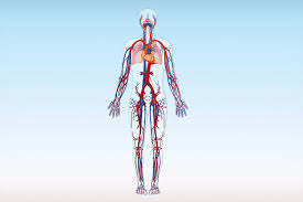
The venous system is the system of veins in the systemic and pulmonary circulations that return blood to the heart.
In the systemic circulation the return is of deoxygenated blood from the organs and tissues of the body, and in the pulmonary circulation the pulmonary veins return oxygenated blood from the lungs to the heart.
When the body is in a supine position, central venous pressure ranges from 8-12 mm Hg and as high as 90 mmHg when the legs are in the standing position.
The venous system comprises superficial and deep veins.
Deep veins are responsible for greater than 90% of the venous return to the heart.
Muscle contraction of the foot and calf initiates cephalad flow of blood.
It opens one way valves within the veins, preventing retrograde flow after closing.
Dysfunctional veins caused venous reflux with backflow of blood, resulting in venous hypertension, and leads to the deliberate delirious effects overtime.
Prolonged venous hypertension causes endothelial dysfunction, increases vascular permeability, and induces vein inflammation.
This process results in disruption of the reduction-oxidation balance, which leads to tissue degradation and clinical manifestations of varicose veins, edema, skin changes, and venous ulcers.
Vein structure consists of three main layers: an outer layer of connective tissue, a middle layer of smooth muscle, and an inner layer lined with endothelium.
Most veins carry deoxygenated blood from the tissues back to the heart; exceptions are those of the pulmonary and fetal circulations which carry oxygenated blood to the heart.
Veins return deoxygenated blood to the heart via the deep veins.
There are three sizes of veins: large, medium, and small.
Smaller veins (venules), and the smallest the post-capillary venules are microscopic that make up the veins of the microcirculation.
Veins are often closer to the skin than arteries.
Veins have less smooth muscle, connective tissue and have wider internal diameters than arteries, allowing them to expand and hold more blood.
Due to thinner walls and wider lumens they are able to expand and hold more blood than arteries.
This capacity is referred to as its capacitance.
Nearly 70% of the total volume of blood in the human body is in the veins.
In medium and large sized veins the flow of blood is maintained by unidirectional venous valves preventing backflow.
In the lower limbs unidirectional venous valves are aided by muscle pumps, also known as venous pumps that exert pressure on intramuscular veins when they contract and drive blood back to the heart.
Veins have a similar three-layered structure to arteries.
The layers have a concentric arrangement that forms the wall of the vessel.
The outer layer, is a thick layer of connective tissue called the tunica externa or adventitia; this layer is absent in the post-capillary venules.
The middle layer, consists of bands of smooth muscle and is known as the tunica media.
The inner layer, is a thin lining of endothelium known as the tunica intima.
The tunica media in the veins is much thinner than that in the arteries as the veins are not subject to the high systolic pressures that the arteries are.
There are valves present in many veins that maintain unidirectional flow.
Unlike arteries, the location of veins varies among individuals.
Veins close to the surface of the skin appear blue due to the alteration of color perception related to the light-scattering properties of the skin and the processing of visual input by the visual cortex, rather than the actual color of the venous blood which is dark red.
Almost 70% of the blood in the body is in the veins, and almost 75% of this blood is in the small veins and venules.
All of the systemic veins are tributaries of the largest veins: the superior and inferior vena cava, which empty the oxygen-depleted blood into the right atrium of the heart.
The thin walled veins, with their greater internal diameters enable them to hold a greater volume of blood, and this greater capacitance gives them the term of capacitance vessels.
It allows for the accommodation of pressure changes in the system.
The whole of the venous system, is a large volume, low pressure system.
The venous system is often asymmetric.
Positions of venae cavae and vessels of the pulmonary circulation Veins vary in size from the smallest post-capillary venules, and more muscular venules, to small veins, medium veins, and large veins.
The thickness of the walls of the veins varies as to their location.
In the legs the vein walls are much thicker than those in the arms.
In the circulatory system, blood first enters the venous system from capillary beds.
Arterial blood changes to venous blood.
Large arteries such as the thoracic aorta, subclavian, femoral and popliteal arteries lie close to a single vein that drains the same region.
Other arteries are often accompanied by a pair of veins held in a connective tissue sheath.
The accompanying veins, known as venae comitantes, run on either side of the artery, and when an associated nerve is also enclosed, the sheath is known as a neurovascular bundle.
This close proximity of the artery to the veins helps in venous return due to the pulsations in the artery.
It also allows for the promotion of heat transfer from the larger arteries to the veins to preserve normal body heat.
The first entry of venous blood is from the convergence of two or more capillaries into a microscopic, post-capillary venule.
Post-capillary venules have a diameter of between 10 and 30 micrometres (μm), and are part of the microcirculation.
Their endothelium is made up of flattened oval shaped cells surrounded by a basal lamina.
Post-capillary venules are too small to have a smooth muscle layer and are instead supported by pericytes that wrap around them.
Post-capillary venules become muscular venules when they reach a diameter of 50 μm, and can reach a diameter of 1 mm.
These larger venules feed into small veins.
The small veins merge to feed as tributaries into medium-sized veins.
The medium veins feed into the large veins which include the internal jugular, and renal veins, and the venae cavae that carry the blood directly into the heart.
The venae cavae enter the right atrium of the heart from above and below.
From above, the superior vena cava carries blood from the arms, head, and chest to the right atrium of the heart, and from below, the inferior vena cava carries blood from the legs and abdomen to the right atrium.
The inferior vena cava is the larger of the two.
The inferior vena cava is retroperitoneal and runs to the right and roughly parallel to the abdominal aorta along the spine.
The three main compartments of the venous system are the deep veins, the superficial veins, and the perforator veins.
Superficial veins are those closer to the surface of the body, and have no corresponding arteries.
Deep veins are deeper in the body and have corresponding arteries.
Perforator veins drain from the superficial to the deep veins.
These are usually referred to in the lower limbs and feet.
Superficial veins include the very small spider veins of between 0.5 and 1 mm diameter, and reticular or feeder veins.
Venous plexuses where veins are grouped or sometimes combined in networks at certain body sites.
The Batson venous plexus, runs through the inner vertebral column connecting the thoracic and pelvic veins.
These veins are noted for being valveless, believed to be the reason for metastasis of certain cancers.
A subcutaneous venous plexus is continuous, and has a high rate of flow is supplied by small arteriovenous anastomoses.
It is the high rate of flow ensures heat transfer to the vein wall.
Blood flows back to the heart in the systemic deep veins.
The flow of blood is maintained by one-way valves in the deep veins, superficial veins, and in the perforator veins.
These venous valves prevent backflow due to the low pressure of veins, and the pull of gravity.
Venous valves also prevent the over-widening of the vein.
Venous valves are bicuspid with two leaflets formed by an infolding of part of the tunica intima.
The leaflets are covered with endothelium, and strengthened with collagen, and elastic fibers.
The endothelial cells on the surfaces of the leaflets are arranged transversely.
On the leaflet surfaces that open to let the blood flow, the cells are arranged longitudinally in the direction of the flow.
Where the leaflets attach, the vein becomes dilated on each side, widening forming hollow cup-shaped pocket regions, known as the valvular sinuses.
The endothelial cells in the sinuses are able to stretch twice as much as those in areas without valves, and when the blood tries to reverse its direction the sinuses fill first closing the leaflets and keeping them together.
Approximately 95% of the venous valves are located in the small veins of less than 300 micrometres.
The deep veins of the lower limb include: the common femoral vein, femoral vein, and the deep femoral vein; the popliteal vein, the tibial, and fibular veins.
The valves also divide the column of blood into segments which helps move the blood unidirectionally to the heart.
Valve action is supported by the action of skeletal muscle pumps that contract and compress the veins.
A skeletal muscle contraction of the muscle makes it wider resulting In compression on the vein that pushes the blood forward.
Valves in the perforating veins close when a calf muscle contracts, to prevent backflow from the deep veins to the superficial.
There are more valves in the lower leg, due to increased gravitational pull, with the number decreasing as the veins travel to the hip.
There are no valves in the veins of the thorax or abdomen.
There is a valve at the junction of the inferior vena cava and the right atrium known as the valve of inferior vena cava also known as the eustachian valve.
This valve is an embryological remnant and is insignificant in the adult.
In the coronary circulation, the blood supply to the heart, is drained by coronary veins that remove the deoxygenated blood from the heart muscle.
These veins include the great cardiac vein, the middle cardiac vein, the small cardiac vein, the smallest cardiac veins, and the anterior cardiac veins.
Cardiac veins carry blood with a poor level of oxygen, from the heart muscle to the right atrium.
Most of the blood of the cardiac veins returns through the coronary sinus.
Generally, the heart veins that go into the coronary sinus: the great cardiac vein, the middle cardiac vein, the small cardiac vein, the posterior vein of the left ventricle, and the oblique vein of the left atrium.
Heart veins that go directly to the right atrium: the anterior cardiac veins, and the smallest cardiac veins (Thebesian veins).
In the bronchial circulation that supplies blood to the lung tissues, bronchial veins drain venous blood from the large main bronchi into the azygous vein, and ultimately the right atrium.
Venous blood from the bronchi inside the lungs drains into the pulmonary veins and empties into the left atrium; since this blood never went through a capillary bed it was never oxygenated and so provides a small amount of shunted deoxygenated blood into the systemic circulation.
In the cerebral circulation supplying the cerebrum the venous drainage can be separated into two subdivisions: superficial and deep.
The superficial system is composed of dural venous sinuses, composed of dura mater walls as opposed to a traditional vein.
The dural sinuses are therefore located on the surface of the cerebrum.
The most prominent dural sinus is the superior sagittal sinus which flows in the sagittal plane under the midline of the cerebral vault, posteriorly and inferiorly to the confluence of sinuses, where the superficial drainage joins with the sinus that primarily drains the deep venous system.
Two transverse sinuses bifurcate and travel laterally and inferiorly in an S-shaped curve that forms the sigmoid sinuses which go on to form the two jugular veins.
In the neck, the jugular veins parallel the carotid arteries and drain blood into the superior vena cava.
The deep venous drainage is primarily composed of traditional veins inside the deep structures of the brain, which join behind the midbrain to form the vein of Galen.
The vein of Galen merges with the inferior sagittal sinus to form the straight sinus which then joins the superficial venous system at the confluence of sinuses.
A portal venous system is a series of veins or venules that directly connect two capillary beds.
The two systems in verebrates are the hepatic portal system, and the hypophyseal portal system.
Circulatory anastomoses, one of which is the join between an artery with a vein known as an arteriovenous anastomosis.
An arteriovenous anastomosis connection is highly muscular, and enables venous blood to travel directly from an artery into a vein without having passed from a capillary bed.
Abnormal connections are known as arteriovenous malformations.
Arteriovenous malformations are usually congenital and the connections are made from a tangle of capillaries.
A small specialized arteriovenous anastomosis known as a glomus body or organ.
A glomus body serves to transfer heat in the fingers and toes.
A glomus body connection is surrounded by a capsule of thickened connective tissue.
There are a great number of glomus bodies in the hands and feet.
A vascular shunt can also bypass the capillary bed and provide a route for blood supply directly to a collecting venule.
The three layers of the vein wall are the outer tunica externa, the middle tunica media and the inner tunica intima.
There are also numerous valves in many of the veins.
The outer tunica externa, also known as the tunica adventitia is a sheath of thick connective tissue.
The middle tunica media is mainly of vascular smooth muscle cells, elastic fibers and collagen.
The middle tunica media layer is much thinner than that in arteries.
Vascular smooth muscle cells control the size of the vein lumens, and thereby help to regulate blood pressure.
The inner tunica intima is a lining of endothelium comprising a single layer of extremely flattened epithelial cells, supported by connective tissue.
The endothelial cells continuously produce nitric oxide a soluble gas, to the cells of the adjacent smooth muscle layer.
The synthesis is carried out by the enzyme endothelial nitric oxide synthase.
Other endothelial secretions are endothelin, and thromboxane, vasoconstrictors and prostacyclin a vasodilator.
In the systemic circulation, veins serve to return oxygen-depleted blood from organs, and tissues to the right heart.
Veins have thinner walls than arteries, and a wider diameter that allow them to expand and hold a greater volume of blood, and provides capacitance that makes possible the accommodation of different pressures in the system.
The venous system apart from the post-capillary venules is a high volume, low pressure system.
The venous system’s Vascular smooth muscle cells control the size of the vein lumens, and thereby help to regulate blood pressure.
The post-capillary venules are exchange vessels with ultra-thin walls that allow the ready diffusion of molecules from the capillaries.
The return of blood to the heart is assisted by the actions of the muscle pump, and by the thoracic pump action of breathing during breathing.
Low venous return can occur from venous pooling with prolonged standing or sitting.
Fainting can occur but usually baroreceptors within the aortic sinuses initiate a baroreflex such that angiotensin II and norepinephrine stimulate vasoconstriction and heart rate increases to return blood flow.
Neurogenic and hypovolemic shock can also cause fainting: the smooth muscles surrounding the veins become slack and the veins fill with the majority of the blood in the body, keeping blood away from the brain and causing unconsciousness.
Jet pilots wear pressurized suits to help maintain their venous return and blood pressure.
Most venous diseases involve obstruction such as a thrombus or insufficiency of the valves, or both of these.
Other venous conditions may be due to inflammation, or compression.
Ageing is a major independent risk factor for venous disorders.
The medical speciality involved with the diagnosis and treatment of venous disorders is known as phlebology.
Venous diseases:
Venous insufficiency is the most common disorder of the venous system, and is usually manifested as either spider veins or varicose veins.
Several treatments for venous insufficiency are available including endovenous thermal ablation using radiofrequency or laser energy, vein stripping, ambulatory phlebectomy, foam sclerotherapy, laser, or compression.
Postphlebitic syndrome is venous insufficiency that develops following deep vein thrombosis.
Venous thrombosis is the formation of a thrombus in a vein.
This most commonly affects a deep vein known as deep vein thrombosis (DVT), but can also affect a superficial vein known as superficial vein thrombosis (SVT).
DVT usually occurs in the veins of the legs, although it can also occur in the deep veins of the arms.
Risk factors: Immobility, active cancer, obesity, traumatic damage and congenital disorders that make clots.
It can cause the affected limb to swell, and cause pain and an overlying skin rash.
A deep vein thrombosis can extend, or a part of a clot can break off as an embolus and lodge in a pulmonary artery in the lungs, known as a pulmonary embolism.
Treatment generally involves anticoagulation to prevents clots or to reduce the size of the clot. Intermittent pneumatic compression is a method used to improve venous circulation in cases of edema or in those at risk from a deep vein thrombosis.
SVT is the development of a thrombus in a superficial vein. SVT is not normally clinically significant, but the thrombus can migrate into the deep venous system where it can also give rise to a pulmonary embolism.
The main risk factor for SVT in the lower limbs is varicose veins.
The portal vein also known as the hepatic portal vein carries blood drained from most of the gastrointestinal tract to the liver.
Portal hypertension is mainly caused by cirrhosis of the liver.
Other causes include an obstructing clot in a hepatic vein (Budd Chiari syndrome) or compression from tumors or tuberculosis lesions
.When the pressure increases in the portal vein, a collateral circulation develops, causing visible veins such as esophageal varices.
Phlebitis is the inflammation of a vein, and usually accompanied by a blood clot when it is known as thrombophlebitis.
When the affected vein is a superficial vein in the leg, it is known as superficial thrombophlebitis, and unlike deep vein thrombosis there is little risk of the clot breaking off as an embolus.
Some disorders as syndromes result from compression of a vein: a venous type of thoracic outlet syndrome, due to compression of a subclavian vein; nutcracker syndrome most usually due to compression of the left renal vein, and May–Thurner syndrome associated with compression of the iliac vein which can lead to iliofemoral DVT, Compression of the superior vena cava most usually by a malignant tumor can lead to superior vena cava syndrome.
A vascular anomaly can be either a vascular tumor or a birthmark, or a vascular malformation.
Infantile hemangioma the vascular mass is soft, easily compressed, and their coloring is due to the dilated anomalous involved veins.
Infantile hemangiomas are most commonly found in the head and neck.
Venous malformations can often extend deeper from their surface appearance, reaching underlying muscle or bone: In the neck they may extend into the lining of the mouth cavity or into the salivary glands.
Venous malformations are the most common of the vascular malformations.
Venous malformation can involve the lymph vessels as a lymphaticovenous malformation.
Venous access is any method used to access the bloodstream through the veins, either to administer intravenous therapy such as medication, or fluid, parenteral nutrition, to obtain blood for analysis, or to provide an access point for blood-based treatments such as dialysis or apheresis.
Ultrasound, particularly duplex ultrasound, is the most usual and widely used way of viewing veins in the diagnosis of venous disease.
Venography is an invasive procedure that uses a catheter to deliver a contrast agent in giving an X-ray of veins.