 Stewart–Bluefarb syndrome is a rare condition involving skin lesions that share clinical features with Kaposi sarcoma and that are secondary to an underlying arteriovenous fistula.
Acroangiodermatitis or pseudo-Kaposi sarcoma are synonymous terms encompassing 2 different conditions: acroangiodermatitis of Mali and Stewart–Bluefarb syndrome.
Their name arises from the clinical and histological similarities both entities share with classic Kaposi sarcoma.
Mainly develops bilaterally on the limbs of elderly patients as a result of chronic venous insufficiency and is an extreme form of stasis dermatitis.
Stewart–Bluefarb syndrome mainly affects the limbs of young patients unilaterally secondary to an underlying arteriovenous malformation.
These structures were distributed diffusely throughout the papillary dermis and formed lobular structures in the reticular dermis, and were accompanied by red-cell extravasation.
Doppler ultrasound revealed an underlying arteriovenous fistula, with low flow resistance and long diastoles in the popliteal artery afferent to the malformation, and an arterialized venous flow in a venous collector.
Stewart–Bluefarb syndrome is an uncommon entity that usually occurs in young patients who present an underlying arteriovenous malformation.
It generally occurs unilaterally on the dorsum of the foot, on the ankle, and on the calf.
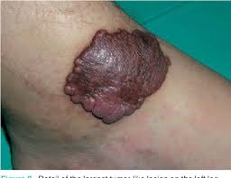
The lesions present as brown or violaceous plaques and macules.
Lesions can grow progressively and develop verrucous lesions and ulcers,with the development of edema, local temperature increase, soft-tissue hypertrophy, varices, stasis changes, and hypertrichosis.
Arteriovenous fistula is suspected by the palpation of a thrill, auscultation of a murmur, or detection of asymmetrical arterial pulses.
Doppler ultrasound is a highly sensitive screening method, and arteriography is the complementary examination of choice in the case of a well-founded suspicion of arteriovenous fistula.
The most typical angiographic finding is early venous filling, which is proportional to the length of the fistula.
Radioisotope scanning is less invasive than arteriography.
The histological findings: proliferation of capillaries in the superficial and middle dermis accompanied by fibroblasts, extravasated red cells, and hemosiderin deposits.
The absence of proliferation of spindle-shaped CD34+ cells and of vascular slits, which are typical of Kaposi sarcoma.
Unlike classic Kaposi sarcoma, human herpes virus type 8 is not detected and the endothelial cells are positive for factor VIII.
The increase in venous pressure resulting from the arteriovenous malformation may stimulate the proliferation of endothelial cells.
An arteriovenous steal syndrome with distal ischemia may induce a local increase in vascular endothelial growth factor, which would lead to endothelial proliferation.
Mast cells proliferate in endothelial and perivascular cells under conditions of ischemia.
Treatment is usually conservative.
Compression, elevation of the limb, and care of ulcers, and infections.
Surgery can lead to ulceration or other complications and only resolves macroscopically detectable fistulas.
Amputation may occasionally be necessary.
Surgery is indicated in cases involving functional impotence, refractory pain, recurrent infection, bleeding, or cardiac decompensation.
Selective embolization may be a valid treatment.