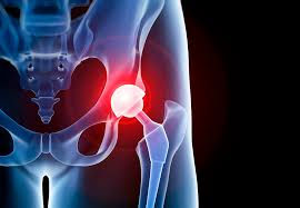
Prosthetic joint infections involve interactions between microorganisms in the implant, and the host immune system.
Microorganisms, typically bacteria, and in rare cases, fungi, adhere to and form biofilms on arthroplasty surfaces.
Biofilms tend to be refractory to many antimicrobial agents and the hosts immune system.
Causative micro organisms are often skin, microbiota inoculated, at placement of the implant: although implants may be seated after placement, either hematogenously or through compromised local tissues.
The most common microorganism group is coagulase negative staphylococci, especially staphylococcus epidermidis, followed by staphylococcus aureus, streptococcus, species, enterococcus species, cutibacterium, species, and Enterobacterales.
As the number of joint replacement increases, the number of joint infections will rise as well.
The incidence of hip and knee PJIs in the United States is about 2.1% and 2.3%, respectively.
The risk of PJI is highest in early postoperative period, but risk persists for the lifetime of the joint, with a significant number of infections manifest after one year.
Studies suggest PJIs are monomicrobial in 70% of cases and 25% are polymicrobial.
Culture negative rates are reported up to 45% of cases.
Cutibacterium acnes accounts for approximately 44% of cases of shoulder PJI.
The risk of prosthetic joint infection is highest in the early postoperative period, but the risk persists for the lifetime of the joint, with a significant proportion of infections, manifesting after one year.
Risk factors include: anemia, injection, drug use, malnutrition, obesity, poor glycemic control, infections, and tobacco use.
Receipt of injections, such as glucocorticoids, hyaluronic acid, or anesthetics into affected joints, three months or less before knee or hip arthroplasty is a risk factor for prosthetic joint infections.
Patients who have undergone multiple arthroplasties and present with prosthetic joint infection in one joint have up to a 20% risk of infection in another joint, either synchronously or metachronously.
Patients with Medicaid, are at increased risk for a PJI.
A prolonged operative time increases the risk of PJI: operative times exceeding 90 minutes increases PJI risk by 1.6, compared to operative times with less than 60 minutes.
Complications to surgery are more likely when arthroplasty is performed at a low volume hospital by low volume surgeons.
Joint replacements most commonly performed in the US are: knee, hip, shoulder, elbow, wrist, ankle, metacarpophalangeal, and interphalangeal joint replacements.
Preoperative screening for it S. aureus carriage, with de colonization of carriers or universal decolonization is considered, and patients undergoing elective surgery should cleanse the skin with chlorhexidine the night before surgery.
Plain x-rays have low sensitivity and low specificity for detecting infection associated with a prosthetic joint.
X-rays may show periprosthetic radiolucency, osteolysis, migration in patients with either infection or aseptic loosening of the prosthesis.
CT and MRI examinations produce artifacts related to the prosthesis. Implants that are not f2242omagnetic, such as titanium or tantalum, are associated with minimal MRI artifact
Bone scans are sensitive for detecting failed implants but are nonspecific for detecting infection.
Bones scans may remain abnormal for more than a year after implantation.
Radioactive labeled leukocyte imaging combined with bone marrow imaging is more accurate than bone imaging alone.
CT scans an MRI may be beneficial, while plane radiographs, have low sensitivity and specificity.
Staphylococci account for more than half of cases of prosthetic hip and prosthetic knee infections (Trampuz).
S. aureus infection is common agent in patients with rheumatoid arthritis and prosthetic joint infections.
Propionibacterium acnes is a common cause of infection with shoulder joint replacement.
About 20% of cases are related polymicrobial infections with most commonly MRSA or anaerobic organisms.
About 70% of prosthetic joint infections are monomicrobial, and 25% polymicrobial.
About 7% of cases cultures are negative and may be related to previous antimicrobial agents:in some studies cultures are negative end up to 45% of cases.
Blood cultures are positive in approximately 25% of cases, most often in acute PJI
Infections of joints involve interactions between the implant, the host’s immune system, and the involved organisms.
Only a small number of microorganisms are needed to seed an implant as they adhere to the implant and form a biofilm protecting them from antimicrobial agents and the host’s immune system.
Microorganisms are often inoculated at joint implantation from the skin, although some organisms can seed the implant hematogenously or through compromised local tissues.
Infection with a virulent organism usually manifests as an acute infection within the first three months after surgery
Infection with less virulent organisms usually manifests as a chronic infection several months or even years postoperatively.
The most common symptom of prosthetic joint infection is pain.
Local signs of swelling, erythema, and warmth at the infected joint site along with fever are common manifestations.
Arthrocentesis is highly recommended for PJI diagnosis.
The volume of aspirate should be at least 3.5 cc for typical microorganisms. The synovial fluid should be assessed for leukocyte, count and neutrophil percentage and for culture.
Additional tests include alpha-defensible, C reactive, protein, leukocyte,esterase, and calprotectin for challenging cases.
Synovial fluid should be cultured aerobically, and anaerobically.
Gram staining is not recommended.
If implant components are removed, culture of implant surfaces, which detects biofilms is useful for microbiological diagnosis
Arthroscopy with biopsy may be considered if no organism is found, the PJI diagnosis is unconfirmed, and surgery is not planned.
Fever can be present in 14% of patients with a chronic prosthetic joints infection but up to 75% of patients if the etiology of a prosthetic joint infection is hematogenous.
Articular, effusion and swelling may be present in 29 to 75% of prosthetic joint infections of the knee, and delayed wound healing, wound dehiscence and wound drainage may accompany up to 44% of joint infections.
The presence of a sinus tract or purulent drainage has a specificity between 97 and 100% as a positive predictive value for prosthetic infection.
Joint stiffness has reported sensitivity of 20% and specificity of 99% in patients with a hematogenous source of a prosthetic joint infection.
Chronic infection may present with pain alone and is often accompanied by loosening of the prosthesis at the bone-cement interface, and sometimes by sinus tract formation.
It may be difficult to differentiate prosthetic joint infection from non-infectious causes of arthroplasty failure.
Arthroplasty procedure should be deferred when there is an active infection elsewhere in the body.
Receipt of injections, such as glucocorticoids, hyaluronic acid, or anesthetics into affected joints, three months or less before knee or hip arthroplasty is a risk factor for prosthetic joint infections.
Surgical-site infection (SSI) after total joint arthroplasty (TJA) continues to pose a challenge and place a substantial burden on patients, surgeons, and the healthcare system.
Estimated 1.0% to 2.5% annual incidence of surgical-site infection after total joint arthroplasty.
Advances in surgical technique, sterile protocol, and operative procedures have minimized SSIs
Preoperative skin preparation have shown varying outcomes after TJA.
Preoperative patient optimization of nutritional status, immune function, and metabolic control are essential to prevent infection.
Intraoperative infection prevention measures include: skin preparation, gloving, surgical drapes, OR staff traffic and ventilation flow, and antibiotic-loaded cement.
Revision procedures for infection after total hip arthroplasty are associated with more hospitalizations, more operations, longer hospital stay, and higher outpatient costs in comparison with primary total hip replacement or revision surgeries for aseptic loosening.
SSI that develops into a periprosthetic infection, can be disastrous.
Infection at the site of a total joint arthroplasty can be classified into four basic categories: Type I, early postoperative, Type II, late chronic, Type III, acute hematogenous, and Type IV, positive intraoperative cultures with clinically unapparent infection.
Treatment: the aim of treatment is to ensure functional, pain-free joints, and cure the infection.
Antibiotic therapy without surgical intervention fails in most cases.
Meticulous surgical debridement is important.
For acute prosthetic joint infections of the hip or knee, debridement, antibiotics, and implant retention may be used, unless a sinus tract is present, the prosthesis is loose, or the wound cannot be closed.
The current standard of care for late chronic infection is considered to be two-stage revision arthroplasty with removal of the prosthesis and cement, debridement, and placement of an antibiotic-impregnated cement spacer, intravenous antibiotics, and a delayed second-stage revision arthroplasty.
chronic infections require resection, arthroplasty, either one stage revision removal of the infected prosthesis, and re-implantation of it new prosthesis during one procedure or two stage revisions.
Antibiotic eluding polymethylmethacrylate articular spaces used into stage revisions help to maintain function during the prosthesis free interval.
Increasing evidence suggests that one stage revision may be acceptable in carefully selected patients.
The use of antibiotic-impregnated cement spacers has improved the outcomes of the treatment of infection associated with total joint arthroplasty.
If patients are not candidates for surgery, anti-microbial, suppression can be attempted, although it is unlikely to cure infection, and so antibiotic treatment is often lifelong.
When unacceptable joint function is expected after surgery, or the infection persists despite surgical efforts, resection or arthroplasty with establishment of pseudoarthrosis for hip infection or amputation for knee infection is sometimes considered.
Total joint replacement complicated by infection has a reported prevalence of 0.5% to 3% and with a higher reported prevalence after total knee arthroplasty than after total hip arthroplasty.
There is also a higher rate of infection after revision hip and knee arthroplasties than after primary hip and knee arthroplasties.
Two-stage revision surgery demonstrated the necessity of removing the implants as well as the cement and of introducing antibiotic therapy for definitive treatment, and is the standard of care for a late chronic infection at the site of a total joint replacement.
Two-stage revision arthroplasty without the use of spacers allows complete removal of foreign materials, with later reimplantation after eradication of the infection, but has disadvantages such as as soft-tissue contractures and joint instability with impaired mobility.
Two-stage revision arthroplasty without the use of a spacer makes reimplantation during the second-stage operation more difficult as a result of arthrofibrosis and the loss of tissue planes.
The two-stage revision for the treatment of infection is associated with a lengthier hospital stay, an increased number of hospitalizations, and increased perioperative morbidity than other non infected revision surgeries.
It is presently standard of care to use antibiotic-impregnated polymethylmethacrylate bone-cement with a chronic infection at the site of a total joint replacement.
Spacers provide direct local delivery of antibiotics.
Spacers preserve patient mobility and facilitate reimplantation surgery.
Type I infections, which are early postoperative infections, both superficial and deep, are defined as wound infections that occur less than four weeks after the primary operation.
Superficial Type-I infections are typically treated with debridement and antibiotic therapy.
Deep Type-I infections are usually treated with replacement of the polyethylene insert, retention of the metal prosthetic components, and intravenous administration of antibiotics.
Late chronic infections (Type II) are defined by their occurrence more than four weeks after the operation.
Late chronic infections (Type II) typically present with worsening pain and loosening of the prosthesis and are usually treated with a two-stage reconstruction.
The standard of treatment for late chronic infections is two-stage revision arthroplasty, which includes placement of an antibiotic-impregnated cement spacer after removal of the prosthesis and thorough débridement, followed by a course of intravenous antibiotics and delayed second-stage revision total joint arthroplasty.
Treatment includes removal of all prosthetic components and bone cement, débridement of necrotic and granulation tissue, placement of an antibiotic-impregnated cement spacer, administration of a course of intravenous antibiotics, and delayed reimplantation arthroplasty when there is no longer evidence of infection.
One-stage exchange arthroplasty also has been used, more commonly in Europe.
In one-stage exchange arthroplasty strict patient selection and use of antibiotic-loaded cement for fixation of the prosthesis are strongly recommended.
Acute hematogenous infections (Type III) are defined by bacteremia and are typically managed with débridement, replacement of the polyethylene insert, and retention of the prosthesis if there is no implant loosening, followed by a course of intravenous antibiotics.
In situations with positive intraoperative cultures (Type IV) within days after revision arthroplasty for the treatment of aseptic loosening are typically managed with intravenous antibiotics and retention of the prosthesis.
There are two types of antibiotic-impregnated cement spacers used in two-stage revisions of total hip and knee arthroplasties: nonarticulating and articulating.
Nonarticulating spacers deliver a high concentration of antibiotics locally and maintain joint space for future revision procedures.
Nonarticulating spacers provide a limited range of motion of the joint.
Nonarticulating spacers can cause quadricep or abductor shortening, scar formation, and bone loss.
Ariculating spacers permit more joint motion and can improve function prior to the second-stage reimplantation surgery.
As a result of improved joint function and decreased scar formation facilitated exposure during reimplantation.
In general, the use of articulating spacers have a better outcome than use of a nonarticulating spacers, which may limit joint motion.
Articulating spacers should be the first option since they appear to provide a better functional outcome.
Antibiotic-impregnated cement spacers can maintain limb length, minimize soft-tissue contracture, facilitate reimplantation, and provide local antibiotic therapy.
Antibiotic-impregnated cement spacers vary in form and function.
Spacers are commercially made, or it may be custom-made in the operating room.
The choice of the spacer is based on many factors, including: the amount of bone loss, the condition of the soft tissues, the need for joint motion, the availability of prefabricated spacers or molding methods, and the selection of the antibiotics.
Spacers may be made of polymethylmethacrylate cement, a cement-coated metal composite or a sterile prosthesis partially coated with antibiotic-impregnated cement.
Spacers with antibiotic-loaded cement deliver high doses of antibiotics at the site of the infection and can achieve local concentrations higher than those achieved with systemic antibiotics alone.
Spacers achieve high concentrations of antibiotics at the site of an infection and can be used to treat infected avascular bone that is isolated from systemic antibiotics while avoiding the potential systemic toxicity that can result from intravenous use.
The cost of a knee revision due to infection is twice that of an aseptic revision and three to four times that of a primary total knee replacement.
The most commonly used antibiotics include tobramycin, gentamicin, vancomycin, and cephalosporins.
These antibiotics can be combined to provide broad-spectrum coverage.
Most periprosthetic infections involve gram-positive organisms such as Staphylococcus aureus and Staphylococcus epidermidis.
If the pathogen and antibiotic sensitivity are clearly identifiable one antibiotic should be used.
When the pathogen is unknown, a combination of antibiotics may be required to completely eradicate the infection.
Vancomycin covers methicillin-resistant Staphylococcus aureus.
Gentamicin covers Enterobacteriaceae and Pseudomonas aeruginosa.
Cefotaxime kills gentamicin-resistant organisms.
If an antibiotic-loaded cement had been used for the primary procedure, bacteria survived a high concentration of that antibiotic and will likely be resistant if the same antibiotic is used in the spacer cement.
Fungal infections are extremely rare at the sites of total joint arthroplasties.
The proper dosage of a specific antibiotic to be used in polymethylmethacrylate bone cement for the treatment of an established infection at the site of a prosthetic joint is not standardized, although impregnation withtwo antibiotics has proven to be superior to the use of a single antibiotic.
Thermostable, water-soluble, susceptibility-directed antibiotics should be used.
The amount of antibiotic introduced is increased, the strength of the cement is reduced.
Hand-mixing of additional antibiotics into antibiotic-impregnated bone cement is possible to increase the antibiotic dosage.
Fungal infections with polymethylmethacrylate bone-cement spacers containing 500 mg of amphotericin B are effective treatments.
The safety of antibiotic-impregnated polymethylmethacrylate bone cement has been well documented.
The forces acting on a hip are about 2.5 times that of body weight but can increase to eight times that with a stumble.
Management of PJI is expensive, resource, intensive, and time consuming.
The cost the revisions for PJI is more than five times as high as the cost of revisions for other reasons.
Prolonged antimicrobial therapy, guided by anti-microbial susceptibility testing is used to treat prosthetic joint infections.
Treatments usually or 6 to 12 weeks of antibiotics, initially intravenously and subsequently orally administered.
Cefazolin administered 60 minutes before incision and fused before tourniquet and inflation is used as an antibiotic prophylaxis.
Prosthetic joint infections are associated with extended hospitalizations, high rates of disability, decreased quality of life, high mortality, as compared with noninfected arthroplasties.
The mean rate of infection eradication after one stage and two stage total knee arthroplasty revisions is 87 and 83%, respectively.
The overall success rate of completion of two stage revisions is less than 50%, with completion rate at 43% for hips and 11% for knees.
Studies from Australia and New Zealand with therapies as noted above had clinical cure with the index prosthesis in place of 74%, 49% and 44% for early, late acute, and chronic prosthetic joint infections, respectively.
Mortality at five years if the hip prosthetic joint infection is 21% in a 10 years is 45%.
For two stage revisions, one-year mortality is 13% for total hip arthroplasties and 9% for total knee arthroplasties.
