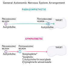
 Pre-ganglionic fibers are part of the autonomic nervous system and originate from the central nervous system (brain or spinal cord) and synapse with post-ganglionic fibers in a ganglion (a cluster of nerve cell bodies outside the central nervous system).
Post-ganglionic fibers then extend from the ganglion to target organs or tissues to carry out the autonomic response.
Provides vasodilation for the coronary vessels of the heart.
Constricts all the intestinal sphincters and the urinary sphincter.
Inhibits peristalsis.
Stimulates orgasm.
Conversely, the PSNS promotes a rest-and-digest response, and promotes the following functions:
Dilates blood vessels leading to the GI tract, increasing blood flow.
Constricts the bronchiolar diameter when the need for oxygen has diminished.
Causes constriction of the pupil and contraction of the ciliary muscle to the lens, allowing for closer vision.
Stimulates salivary gland secretion, and accelerates peristalsis.
Stimulates sexual arousal.
Control of Autonomic Nervous System Function
The medulla oblongata, in the lower half of the brainstem, is the control center of the autonomic nervous system.
LEARNING OBJECTIVES
Describe the control of the autonomic nervous system
KEY TAKEAWAYS
Key Points
The medulla contains the cardiac, respiratory, and vasomotor centers.
The ANS is classically divided into two subdivisions, the sympathetic division and the parasympathetic division.
As a rule, the SNS functions in actions that require quick responses, while the PSNS is initiated in actions that don’t require immediate response.
Key Terms
fight or flight: This theory states that animals react to threats with a general discharge of the sympathetic nervous system, priming the animal for fighting or fleeing.
The autonomic nervous system (ANS) is the part of the peripheral nervous system that controls involuntary functions that are critical for survival. The ANS participates in the regulation of heart rate, digestion, respiratory rate, pupil dilation, and sexual arousal, among other bodily processes.
Within the brain, the ANS is located in the medulla oblongata in the lower brainstem. The medulla’s main functions are to control the cardiac, respiratory, and vasomotor centers, to mediate autonomic, involuntary functions, such as breathing, heart rate, and blood pressure, and to regulate reflex actions such as coughing, sneezing, vomiting, and swallowing.
This is a drawing of the brain and spinal cord. It shows, from top to bottom, the positions of the pineal gland, pituitary gland, pons, cerebellum, and the medulla oblongata.
The brain stem with pituitary and pineal glands: The medulla is a subregion of the brainstem and is a major control center for the autonomic nervous system.
The hypothalamus acts to integrate autonomic functions and receives autonomic regulatory feedback from the limbic system to do so. The ANS is classically divided into two subdivisions, the sympathetic division and the parasympathetic division.
The sympathetic division of the ANS is often referred to as the sympathetic nervous system (SNS). The SNS provides noradrenergic drive to the ANS. It is often referred to as a quick response mobilizing system that initiates the body’s fight-or-flight response.
PSNS input to the ANS is responsible for the stimulation of feed-and-breed and rest-and-digest responses, as opposed to the fight-or-flight response initiated by the SNS. The parasympathetic division of the ANS (PSNS) acts to complement and modulate the drive provided by SNS neurotransmission within the ANS.
As a rule, the SNS functions in actions require quick responses while the PSNS is initiated in actions that don’t require immediate response.